 If you’re experiencing pain in your muscles or bones, your doctor may have ordered a CT scan. A computed tomography (CT) scan is a valuable imaging tool that allows doctors to make decisions quickly. This is critical to diagnose conditions that need urgent care like bone or muscle disorders.
As a patient, you may be wondering what it’s like to get a CT scan for muscle or bone disorders and how your doctor can form a diagnosis based on the scan. In this post, we will answer questions about muscle or bone CT scans to help you decide if it’s right for you. We understand getting a CT scan can be intimidating, but we are here to help you every step of the way. If you have any questions, please reach out to us at Envision Imaging.
If you’re experiencing pain in your muscles or bones, your doctor may have ordered a CT scan. A computed tomography (CT) scan is a valuable imaging tool that allows doctors to make decisions quickly. This is critical to diagnose conditions that need urgent care like bone or muscle disorders.
As a patient, you may be wondering what it’s like to get a CT scan for muscle or bone disorders and how your doctor can form a diagnosis based on the scan. In this post, we will answer questions about muscle or bone CT scans to help you decide if it’s right for you. We understand getting a CT scan can be intimidating, but we are here to help you every step of the way. If you have any questions, please reach out to us at Envision Imaging.
What Is a CT Scan for Muscles and Bones?
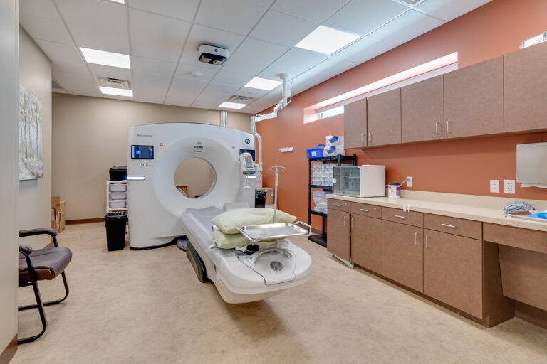
A CT scan is a painless, noninvasive imaging procedure. A CT scan uses x-rays and computer technology to produce highly detailed pictures of the body, including the organs, muscles, bones and fat. These images appear in “slices” so doctors can thoroughly examine the inside of the body without having to do surgery.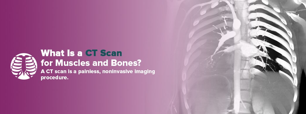 During a regular x-ray, a beam of energy passes through the part of the body needing to be studied. Patients stand in front of an x-ray detector which captures variations of the energy beam after it moves through the body. This forms an image which shows “shadows” produced by objects inside the body. X-rays are helpful for information about bones but may not provide enough information about muscles. However, you can see muscles on a CT scan much better. Doctors may order a CT scan when they need greater detail regarding bones, muscles or both.
A CT scan works like a regular x-ray, except an x-ray beam will move in a circle around your body. This produces many different angles of the area in need of a diagnosis. The x-ray information then goes to a computer which interprets the data and turns it into detailed cross-sectional images. Doctors can look at CT scan images to see the position, size and shape of muscles, bones and organs. A CT scan shows muscle damage and bone abnormalities. You can get a muscle or bone CT scan on any area of your body.
Your doctor may request you to get a CT scan with or without an iodine-based contrast. Contrast is a substance given to you by mouth or through an IV which causes the muscle tissue to show up more clearly on the scan.
During a regular x-ray, a beam of energy passes through the part of the body needing to be studied. Patients stand in front of an x-ray detector which captures variations of the energy beam after it moves through the body. This forms an image which shows “shadows” produced by objects inside the body. X-rays are helpful for information about bones but may not provide enough information about muscles. However, you can see muscles on a CT scan much better. Doctors may order a CT scan when they need greater detail regarding bones, muscles or both.
A CT scan works like a regular x-ray, except an x-ray beam will move in a circle around your body. This produces many different angles of the area in need of a diagnosis. The x-ray information then goes to a computer which interprets the data and turns it into detailed cross-sectional images. Doctors can look at CT scan images to see the position, size and shape of muscles, bones and organs. A CT scan shows muscle damage and bone abnormalities. You can get a muscle or bone CT scan on any area of your body.
Your doctor may request you to get a CT scan with or without an iodine-based contrast. Contrast is a substance given to you by mouth or through an IV which causes the muscle tissue to show up more clearly on the scan.
Schedule a CT Scan at Envision Imaging
How Can CT Scans Be Used to Diagnose Muscle and Bone Disorders?
A CT scan takes detailed images of your musculoskeletal system. Your musculoskeletal system is composed of your muscles, bones, joints and ligaments. Any of these parts could become injured or affected by a disorder. Some disorders, like fractures, mainly affect the bones. Other disorders, like tendinitis, mostly affect muscles and soft tissues. Current CT scan technology allows a doctor to view scans of bones and muscles individually in two-dimensional “slices,” or as a whole three-dimensional image. Doctors can examine these images and quickly identify a wide range of musculoskeletal injuries or conditions such as:- Bone damage, lesions or fractures
- Muscle damage
- Tumors, infections or blood clots
- Internal bleeding
1. CT Scan for Bones
A CT bone scan may help your doctor identify bone disorders such as:- Osteoporosis
- Osteopenia
- Traumatic fractures
- Paget’s disease
- Bone cancer
- Bone infections
2. CT Scan for Muscles
Doctors can use a CT scan to detect muscle damage or any of the following conditions:- Sarcopenia
- Muscular dystrophy
- Myositis
- Cancer
- Muscle sprains or strains
- Tendinitis
Why Should You Consider a CT Scan to Detect Muscle or Bone Damage?
You might need a CT scan if other tests, such as x-rays or MRIs, do not provide enough information. If your doctor recommends a CT scan for a muscle or bone issue, it’s certainly worth weighing the pros and cons. Muscle or bone pain can be debilitating. For example, according to the World Health Organization (WHO), musculoskeletal disorders are the second largest reason for disability worldwide, with lower back pain being the leading cause. There are more than 150 diagnoses that affect the muscles, bones, joints and other parts of the musculoskeletal system. Your muscles and bones help you move your body and keep your shape. It’s critical to your health and well-being to get muscle or bone disorders diagnosed and treated. Overall, CT scans help your doctor provide the best care possible. Your doctor might recommend a CT scan over other forms of diagnostic imaging because CT scans:- Are fast and accurate.
- Provide one of the best methods for detecting cancer.
- Allow doctors to detect injuries after trauma quickly.
- Help doctors evaluate the results of recent surgeries.
- Allow doctors to look at bones, muscle tissue and blood vessels all at once.
- Getting a CT scan is a painless, noninvasive procedure.
- Getting a CT scan is a fast and simple procedure.
- You can get a CT scan regardless of implanted medical devices, unlike an MRI.
- A CT scan may eliminate the need for exploratory surgery or biopsies.
- Radiation leaves the body after a CT scan examination.
- A CT scan machine is less sensitive to movement than an MRI machine.
- A CT scan machine is less likely to trigger claustrophobia than an MRI machine.
What Are the Risks of CT Scans?
Like x-ray machines, CT scanners pass ionizing radiation through the body to create images. This means that the radiation can change the cells in your body as it passes through. CT scanners and x-ray machines use low doses of radiation and cells can usually repair themselves.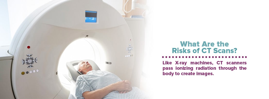 No conclusive evidence proves small amounts of CT scan radiation can cause cancer. However, other studies have shown that large amounts of radiation slightly increase the risk of cancer. Even so, the risk of developing cancer from a CT scan is still very small compared to the average risk. For example, the Food and Drug Administration (FDA) states that there’s about a one in 2,000 chance of getting cancer from a CT scan. For perspective, about one in five people will develop cancer naturally.
The risk of developing cancer from radiation is higher for children than it is for adults. This is because children are still growing and have cells that are more sensitive than older adults. Children also have more life to live which may be enough time for potential effects to show up. CT scan technologists adjust the radiation dose to the patient’s size, so children receive smaller doses than adults.
It’s understandable if you’re worried about exposure to radiation due to cancer risks. However, know that your doctor ordered a CT scan because they felt the benefits outweighed the risks. If you feel uneasy about getting a CT scan, consider expressing your concerns to your doctor. Your doctor may be able to recommend an alternative diagnostic tool that makes you feel more comfortable.
With that said, it’s always good to be aware of the risks with any medical procedure. Here are a few safety notes to keep in mind:
No conclusive evidence proves small amounts of CT scan radiation can cause cancer. However, other studies have shown that large amounts of radiation slightly increase the risk of cancer. Even so, the risk of developing cancer from a CT scan is still very small compared to the average risk. For example, the Food and Drug Administration (FDA) states that there’s about a one in 2,000 chance of getting cancer from a CT scan. For perspective, about one in five people will develop cancer naturally.
The risk of developing cancer from radiation is higher for children than it is for adults. This is because children are still growing and have cells that are more sensitive than older adults. Children also have more life to live which may be enough time for potential effects to show up. CT scan technologists adjust the radiation dose to the patient’s size, so children receive smaller doses than adults.
It’s understandable if you’re worried about exposure to radiation due to cancer risks. However, know that your doctor ordered a CT scan because they felt the benefits outweighed the risks. If you feel uneasy about getting a CT scan, consider expressing your concerns to your doctor. Your doctor may be able to recommend an alternative diagnostic tool that makes you feel more comfortable.
With that said, it’s always good to be aware of the risks with any medical procedure. Here are a few safety notes to keep in mind:
- Generally, CT scans are not recommended for women who are pregnant to avoid potential risk to the fetus.
- Children should only get a CT scan if it is medically necessary.
- Although extremely rare, a contrast agent may cause an allergic reaction in some individuals.
- Patients with kidney problems should speak with their doctor before getting a CT scan with contrast.
How to Prepare for a Muscle and Bone CT Scan
Your doctor should provide instructions on how to prepare for your CT scan. Generally, you can expect the following steps:- Inform your doctor about any allergies, medications, recent illnesses or other medical conditions before your exam.
- Wear comfortable clothing to your exam and expect to change into a hospital gown before the scan.
- Remove metal objects such as jewelry, body piercings, removable dental work, glasses and hairpins before the exam as these may affect the quality of CT scan images.
- If you will be given a contrast agent, you may be asked not to eat or drink a few hours before the exam.
- If you are getting a CT scan without a contrast agent, you can eat and drink as you normally would before the exam.
- You can take your medication as usual before the exam with or without contrast.
What to Expect During the CT Scan
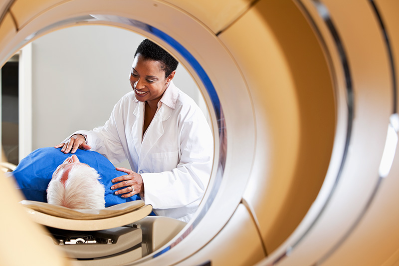
CT scans often take place in an outpatient facility. Sometimes patients receive CT scans in the hospital. If you feel nervous or anxious about getting a CT scan, speak with your doctor ahead of time. They may schedule an anti-anxiety medication to help you feel calmer during the exam.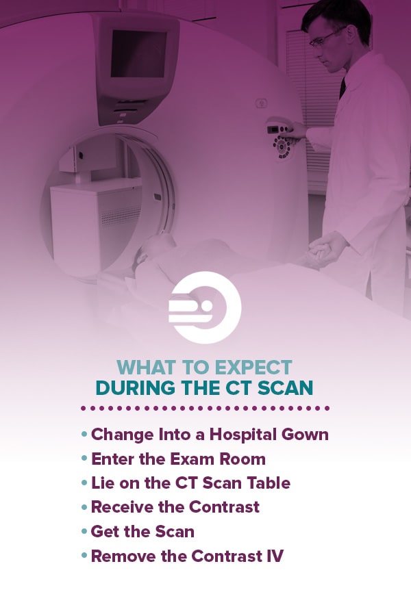 Your procedure will vary depending on your needs and condition. However, here’s what you can generally expect with a CT scan.
1. Change Into a Hospital Gown
You may be asked to change into medical scrubs (top and pants) or a hospital gown when it’s time for the scan. You can store jewelry and other personal belongings in a lockable storage area. It’s best to leave jewelry and valuables at home.2. Enter the Exam Room
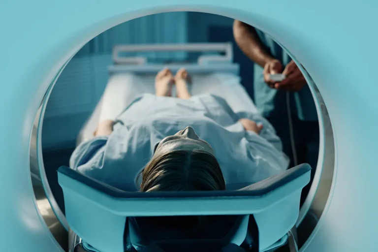
A technologist will guide you to the exam room. They will explain the procedure, and you’ll have a chance to ask questions. You’ll see the CT scanner when you enter the room. Typically, a CT scan machine looks like a donut-shaped tunnel with a table in its center.3. Lie on the CT Scan Table
The technologist will help you onto the table. You will lie flat on the table which will slide in and out of the tunnel. A technologist may arrange pillows or straps to help you feel comfortable and stay still during the scan.4. Receive the Contrast
Depending on the part of the body being scanned, you may be given a contrast agent. The most common type of contrast is a double contrast. With a double contrast, you will be asked to drink a flavored mixture before the exam, and you will be given contrast through an IV once you are in the exam room. The contrast will highlight certain parts of your body to help the doctor see muscles and other tissues more clearly. You may experience a warm feeling throughout your body once you’re given the contrast. Some patients also experience a metallic taste after receiving the contrast. The effects from the contrast only last for a minute or two.5. Get the Scan
The technologist will move to another room where the controls are located. They will be able to see you the whole time through a window and will communicate with you through a two-way microphone. You may also have a call button you can press if you have any issues during the exam. During the exam, you can relax as you try to lie still. You will not feel any pain during the exam. However, you might feel some discomfort from lying still for the duration of the scan, especially if you recently suffered an injury. The technologist will do everything they can to complete the procedure quickly and make you feel as comfortable as possible. Once the scan starts, the scanner will rotate around you, and x-rays will pass through your body for a short amount of time. You may be asked to hold your breath for a few seconds at a time to help reduce movement. You’ll hear whirring and clicking noises during the exam – this is normal. The scanner will detect the x-rays absorbed by your body and send the information to a computer. The computer will then translate the data so a radiologist can read it. A radiologist is a doctor who had been trained to interpret CT scan images. The radiologist will discuss the CT scan results with your doctor.6. Remove the Contrast IV
After the scan, the technologist will remove the contrast IV and help you off the table. You can resume your day as usual unless your doctor says otherwise. A CT scan usually takes 30 minutes to complete.Schedule Your Appointment at Envision Imaging
It’s estimated that over 70 million CT scans are performed each year. CT scans help doctors make critical decisions about their patients’ health. If you’ve decided to go forward with a CT scan for muscle or bone pain, we’re here to help you get through the experience easily and comfortably. At Envision Imaging, we understand how stressful medical testing can be, especially if you’re not feeling well. That’s why we strive to welcome you with warmth and compassion when it’s time for your CT scan. We also use the latest technology to provide fast turnaround rates and accurate results. Our goal is to help your doctor help you, so you can focus on feeling better. To schedule a CT scan appointment, contact us today. Sources:
- https://www.merckmanuals.com/home/bone,-joint,-and-muscle-disorders/symptoms-of-musculoskeletal-disorders/musculoskeletal-pain
- https://www.envrad.com/services/ct-scans/
- https://www.health.harvard.edu/staying-healthy/do-ct-scans-cause-cancer
- https://www.nibib.nih.gov/science-education/science-topics/x-rays
- https://www.hopkinsmedicine.org/health/treatment-tests-and-therapies/computed-tomography-ct-or-cat-scan-of-the-bones
- https://orthoinfo.aaos.org/en/treatment/x-rays-ct-scans-and-mris/
- https://www.webmd.com/cancer/what-is-a-ct-scan#1
- https://www.merckmanuals.com/home/bone,-joint,-and-muscle-disorders/diagnosis-of-musculoskeletal-disorders/introduction-to-diagnosis-of-musculoskeletal-disorders
- https://www.merckmanuals.com/home/bone,-joint,-and-muscle-disorders/diagnosis-of-musculoskeletal-disorders/tests-for-musculoskeletal-disorders
- https://www.radiologyinfo.org/en/info.cfm?pg=bodyct
- https://www.urmc.rochester.edu/encyclopedia/content.aspx?contenttypeid=92&contentid=P07649
- https://www.mayoclinic.org/tests-procedures/ct-scan/about/pac-20393675
- https://www.who.int/news-room/fact-sheets/detail/musculoskeletal-conditions
- https://medlineplus.gov/muscledisorders.html
- https://medlineplus.gov/bonediseases.html
- https://www.radiologyinfo.org/en/info.cfm?pg=about-mission
- https://www.radiologyinfo.org/en/info.cfm?pg=bodyct
- https://www.mayo.edu/research/documents/radiation-exposure-during-imaging-examspdf/DOC-10027821
- https://www.radiologyinfo.org/en/info.cfm?pg=safety-hiw_08
- https://www.cancer.gov/about-cancer/diagnosis-staging/ct-scans-fact-sheet
- https://www.fda.gov/Radiation-EmittingProducts/RadiationEmittingProductsandProcedures/MedicalImaging/MedicalX-Rays/ucm115329.htm
- https://www.envrad.com/locations/
- https://www.semc.org/services-directory/imaging-radiology/diagnostic-imaging-center/computer-tomography-ct-scanning/head-temporal-bone-ct-scan
- https://www.envrad.com/services/x-ray/
- https://www.envrad.com/services/mri-scans/
- https://www.envrad.com/about-us/why-choose-us/