MRI Scans

An MRI Scan (magnetic resonance imaging) is a painless technique that allows doctors to look at the soft tissues of the body.

Jump Ahead:

This scan is used to gather important information about your brain, spine, joints or other internal organs. It helps study nerves, muscles, ligaments, bones, and other tissues. These studies are used to evaluate the possible presence of injuries to these areas. MRIs use magnetic fields and radio waves to obtain incredibly detailed images of the human body.

MRI FAQ's

What do I do if I am pregnant? Is MRI safe?

Currently, we scan pregnant patients only when it is considered medically beneficial. Your referring physician and an MRI Board Certified Radiologist will consult with one another and will sign consent forms deeming the MRI as a medically necessary procedure. Your doctor will then discuss the risks and benefits, and you will be asked to sign a consent form at the time of your appointment. The Radiologist will be available to discuss your exam and the risks and benefits of the procedure with you at the time of your appointment if you would desire.

What can I expect before my MRI exam?

Before your MRI exam, you will fill out a screening form asking about anything that might create a health risk or interfere with the imaging. The screening form will be reviewed by your technologist prior to entering the MR suite.

To ensure your safety during your MRI exam, all patients will be asked to change into MR safe appropriate attire (i.e., a gown or scrub suit) provided by the facility. Your technologist will advise you to remove all metallic objects when changing into the approved garments. This includes hearing aids, hairpins, and all jewelry, to include body piercings when possible. Lockers are available to secure your personal items.

Please consult the MRI technologist if you have any questions BEFORE you enter the MR suite.

What is MRI?

MRI or Magnetic Resonance Imaging is a way to look inside your body without the use of x-rays. It is completely painless. MRI can allow your doctor to see certain types of tissue and can provide very important information about the brain, spine, joints and internal organs. MRI can allow your physician the opportunity for early detection of disease or injuries so proper treatment may be started as soon as possible.

How does MRI work?

Your body is composed of atoms. Water or hydrogen atoms make up 95% of the human body. Usually the hydrogen atoms within the body spin at random. When you have an MRI, you are placed in a strong magnetic field that is up to 8,000 times stronger than that of the earth, which causes these atoms to realign and spin all in the same direction. Like CT, MRI acquires images that are a “slice” of anatomy. Using the magnetic fields and radio waves, remarkably detailed cross-sectional images of the body can be obtained. A computer processes these images to produce detailed pictures of the anatomy.

Can anyone have an MRI?

Because some metals interfere with the function of the MRI equipment, certain patients are not able to have an MRI exam. The following equipment or conditions may create problems with an MRI. Please call with concerns about any of the following metals in your body.

  • A pacemaker or pacing wires
  • Metal fragments in one or both eyes
  • Inner ear implants
  • Cerebral aneurysm clips
  • Implanted neurostimulator
  • Tens unit
  • Certain metal implants

How long does it take?

The exam usually takes between 30 and 60 minutes. If your doctor orders your MRI exam with contrast, the exam may take longer to complete.

What is contrast?

In some cases, your doctor may order your test with contrast. This is a fluid that is injected into a vein (usually in your arm). This helps to make certain details on the exam clearer and is routine for certain MRI exams.

What will happen during the MRI exam?

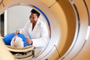
You will be asked to lie down on the examination table on your back. The table will slide smoothly into the opening, and you will be positioned either head first or feet first, depending on the type of exam. Once the exam begins, it is important that you are as still as possible. You will hear “knocking” and “banging” noises, and the hum of the machine. This is all normal and you will be able to talk to the technologist during your procedure. You will be able to listen to the radio, or you may bring a favorite CD, or choose one at our facility.

What if I feel anxious or claustrophobic?

One of the first things we recommend to anyone who thinks they might feel anxious or claustrophobic during an exam is an advanced trip to our facility to actually look at the scanner. We often find that once patients see how wide the opening is and how short the scanner is, their anxiety is eliminated. Remember, MRI scanners have changed dramatically over the last 10 years, and are no longer made with such small, restrictive openings and long “tubes” or “tunnels”.

Our technologists are very skilled at helping you feel relaxed and comfortable during your exam. It often helps to listen to music during your exam. You may have a family member in the room with you if you desire. In very rare instances, a patient may require sedation, which must be scheduled ahead.

Where can I get an MRI done?

If your doctor recommends this type of imaging procedure, you may be wondering where to get your MRI done. Under the umbrella of Envision Radiology, we’re proud to offer MRI exams at many of our locations in Texas, Oklahoma, Louisiana and Colorado. Because each of our locations is locally operated, be sure to check with your office of choice to make sure that MRI imaging services are provided there. You can also click on their individual location page and check out the “Services Provided” section for a full list of offered procedures.

While there are many places that do MRI, none match our level of professionalism, comfort and compassionate patient care. The moment you walk through our door, you’ll be made to feel at ease and walked through the process from beginning to end. Our high-quality services and technological advances ensure that your MRI scans are of the highest quality, and we’ll deliver your results to your referring physician as soon as possible.

Find Your Nearest Location and the Exact Service You Need

Please use our locations page to find your nearest location. Be sure to use the service filter on the left-hand side of the page to find the location/service you are looking for:

Find Your Nearest Location

MRI Short Bore Scans – High-Field (1.5T)

MRI Short Bore scanners offer a comfortable MRI experience.

Short Bore Systems

Short bore systems are 50% shorter and 5% wider than conventional MRI setups. These dimensions allow for a very airy and roomy imaging experience.

High-Field 1.5T

High-Field 1.5T refers to the quality of the images provided. With a wide variety of coil options, 1.5T allows for the potential of better image quality across a variety of imaging applications (in comparison to the 3T) This unit has a well-proven track record as being the best for high-quality images.

MRI Short Bore Scans tend to last 30 to 60 minutes.

MRI Wide Bore Scans – High-Field (1.5T)

Wide Bore Scans

The wide bore MRI creates a wider opening, allowing for a much more comfortable MRI. It differs from the traditional MRI scanner in the size of the opening (“bore”). In a traditional bore, the opening is just slightly larger than the patient, which can tend to create a very restrictive and uncomfortable environment.

High-Field 1.5T

This system is combined with the very powerful 1.5T magnet, creating incredibly high-quality images while maintaining a comfortable patient experience. Compared to the 3T, the 1.5T is well proven as being the best for high-quality imaging.

MRI Wide Bore Scans usually last between 30 and 60 minutes.

MRI Scans – High-Field (1.5T)

The High-Field 1.5T scanner provides the latest in advanced imaging technology.

This particular scanner is considered the industry standard and is valuable for imaging all parts of the body.

This traditional MRI machine with the incredibly powerful High-Field 1.5T magnet takes about 30 to 60 minutes from start to finish.

3T MRI Scans

A 3T MRI scan, also known as a 3 Tesla MRI, is a powerful and efficient imaging test that some patients might have instead of a traditional 1.5T MRI. Though 3T scanners used to be found primarily in medical research centers, you can see them hard at work in clinical settings, as well.

As the name suggests, 3T scanners use strong magnets that create a magnetic field twice as powerful as a 1.5T scanner. As a result, 3T MRIs can produce clearer images in a shorter amount of time.

In comparison to other MRI scans, 3T scans provide more detailed information about the organs and soft tissue. Your doctor may recommend a 3T MRI in order to obtain images of the brain, spine or soft joint tissue, or to see detail inside the bones and blood vessels. 3T MRI scans may also serve as replacements for more invasive vascular studies.

3T can also help doctors differentiate between healthy and diseased tissues more easily, which allows them to provide their patients with earlier, more confident diagnoses. This type of MRI scan might help you if the results of previous imaging tests haven’t provided enough detail to reveal a problem.

Though 3T scans use stronger magnets than typical MRIs, they remain comfortable for the patient. By offering shorter scan times without sacrificing image quality, patients who receive 3T MRIs have a more pleasant test experience overall.

Open MRI Scans

patient entering mri machine

Please note: Open MRI only available at our Envision Imaging of Tulsa location.

An “Open” MRI simply refers to the way the equipment is configured. An Open MRI is open on three sides, creating a very roomy, bright imaging experience. This MRI provides a modern design, yielding fast, comfortable exams.

MRCP Scans

The MRCP scan (magnetic resonance cholangiopancreatography) is a special form of MRI, focused on attaining images of the hepatobiliary and pancreatic systems, including the gallbladder, liver, bile ducts, pancreas, and pancreatic duct.

This is an absolutely painless, non-invasive medical test used to help diagnose abdominal pain, or to evaluate pancreatitis to detect the underlying cause. It also aids in the examination of diseases of the liver, gallbladder, bile ducts, pancreas, and pancreatic duct.

The actual MRCP exam takes about 10 minutes, but it is most often performed with a standard MRI of the abdomen, which may last approximately 30 minutes. In most cases, the entire examination is completed within 45 minutes in total.

MRI Spectroscopy

MRI Spectroscopy is a completely noninvasive technique used to characterize the biochemistry of tumors, infarcts and other pathology.

An MRI Spectroscopy is often done to diagnose certain metabolic disorders, including those affecting the brain. It can help determine the specifics of a tumor, such as a tumor’s aggressiveness, or tumor metabolism.

The MRI Spectroscopy provides additional information on chemicals present in the body’s cells and may be performed during your MRI exam while adding only approximately 15 minutes to the exam time.

MR Elastography

MR Elastography (MRE) is a new way to take images of the body. The scan works by combining MRI imaging with sound waves which help to create a visual map (known as an elastogram). The elastogram can show the stiffness of various body tissues. MRE can be used to detect hardening of the liver caused by numerous types of chronic liver disease.

Schedule an Appointment

If you’re looking for places to get an MRI, schedule an appointment for an MRI scan at one of our offices. Please contact your specific center.