 Early detection and diagnosis of cancer can significantly increase your chances of being treated successfully. Early cancer treatments can also be less expensive and complicated than more advanced disease treatments, which may spare you and your family heartaches.
Early detection also helps doctors identify precancerous tissue abnormalities destined to become life-threatening cancers, which provides the chance for earlier intervention for preventing cancer from ever developing at all. Similarly, being able to identify precancerous tissue abnormalities accurately, and early cancers before they turn into possibly fatal malignancies, can also spare you and your family from the potential financial and physical burdens of unnecessary treatment.
Still, too many individuals are receiving cancer diagnoses at late stages, jeopardizing their chances of successful treatment and long-term survival. Imaging in cancer diagnosis can be a beneficial tool to detect malignancies at their earliest stages to increase your treatment success.
Early detection and diagnosis of cancer can significantly increase your chances of being treated successfully. Early cancer treatments can also be less expensive and complicated than more advanced disease treatments, which may spare you and your family heartaches.
Early detection also helps doctors identify precancerous tissue abnormalities destined to become life-threatening cancers, which provides the chance for earlier intervention for preventing cancer from ever developing at all. Similarly, being able to identify precancerous tissue abnormalities accurately, and early cancers before they turn into possibly fatal malignancies, can also spare you and your family from the potential financial and physical burdens of unnecessary treatment.
Still, too many individuals are receiving cancer diagnoses at late stages, jeopardizing their chances of successful treatment and long-term survival. Imaging in cancer diagnosis can be a beneficial tool to detect malignancies at their earliest stages to increase your treatment success.
What Are Imaging Tests, and How Can They Detect Cancer?
Imaging tests help doctors take a look at what’s occurring inside your body, and can also show the doctor how far your cancer has spread, and whether the treatment is working. Imaging tests work by sending types of energy — like sound waves, x-rays, magnetic fields or radioactive particles — through your body. The tissues of your body then change these energy patterns to create a picture or image. Doctors use imaging tests for cancer in various ways, including:- Identifying cancer in its earliest stages when it’s small and hasn’t spread, and you’re not experiencing symptoms.
- Looking for a lump or tumor if you are experiencing symptoms. They can also see if it’s cancer or another disease causing your symptoms.
- Predicting if your tumor is potentially cancerous and whether the doctor needs to remove and analyze a small tissue sample to help determine this. Usually, doctors can use the biopsy process to see if any change is cancer.
- Seeing where your tumor sits, even if it’s deep inside your body.
- Finding out what stage your cancer is in, or if it has spread to other parts of your body.
- Planning treatment, such as showing where the doctor needs to target cancer radiology therapy beams.
- Detecting if your tumor has grown, shrunk or stayed the same following treatment. This lets the doctor know how well your treatment is working, and whether any changes in therapy are necessary.
- Identifying if your cancer returned after treatment.
Schedule an Appointment at Envision Imaging
Types of Imaging Tests to Detect Cancer and the Benefits of Each
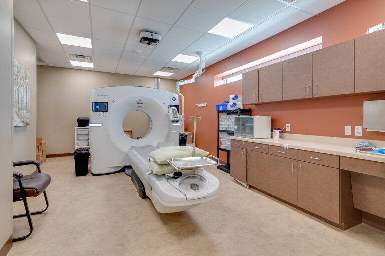
So, what tests are done to check for cancer? Different types of scans for cancer include the following.1. Computed Tomography (CT) Scan
The doctor uses a CT scan to take images of the inside of your body from various angles using x-rays. Then, a computer combines the pictures into a three-dimensional, detailed image to reveal any tumors or abnormalities. In some cases, the doctor will order a specialized dye called a contrast medium, which the technician will give you before your scan to provide better image detail. They’ll either inject the dye into your vein or provide a liquid or pill you swallow.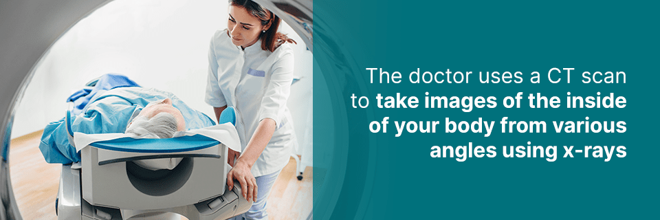 Common scanned areas include the:
- Neck
- Head
- Abdomen
- Chest
- Limbs
- Pelvis
Benefits of a CT Scan
CT scans can help doctors:- Learn the stage of your cancer. When the doctor knows this, they can choose the best course of treatment and maybe even predict your chances of recovery.
- Identify the correct area for a biopsy.
- Plan radiation therapy.
- Assess the effectiveness of treatment during follow-up visits.
- Assess the success of treatment after its completion and during follow-up care.
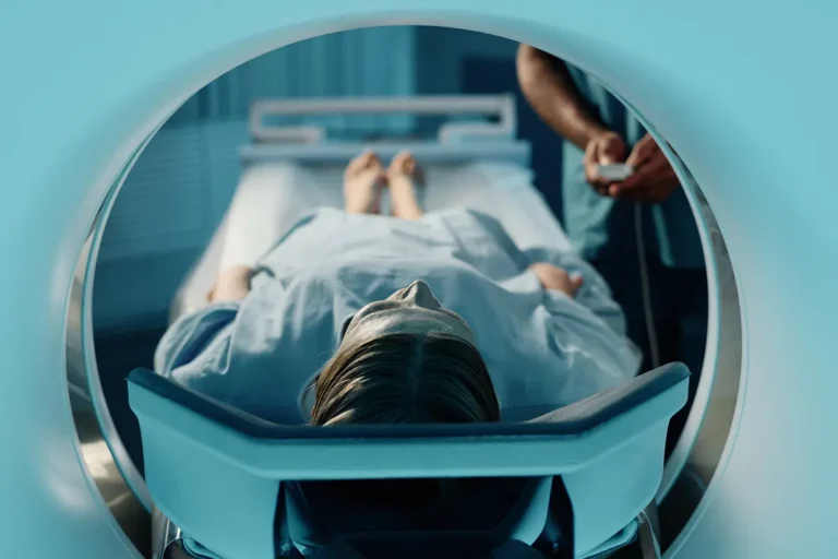
2. Magnetic Resonance Imaging (MRI)
An MRI scan uses powerful radio waves and magnets to generate detailed, computer-generated images of your body. Doctors also use it to measure the size of your tumor. There’s a narrow, tunnel-like opening on a standard MRI machine. Some centers have more “open” or less confining MRI machines, which are ideal for patients who have claustrophobic tendencies. An MRI doesn’t use x-rays or other types of radiation. Because of this, physicians seek the help of an MRI to look for issues in the male and female reproductive systems. It’s typically safe, even if you’re pregnant. Doctors use MRIs to take pictures of your spinal column, brain, chest, abdomen and breast.Benefits of an MRI
MRIs can help doctors:- Identify if a tumor is cancerous or noncancerous.
- Determine the size and location of a tumor.
- Monitor the effectiveness of treatment.
- Plan cancer treatment, such as radiation therapy or surgery.
3. Ultrasound
Ultrasounds are imaging tests, also referred to as ultrasonography or sonography. An ultrasound creates images of internal organs using high-frequency sound waves. These sound waves hit your organs, bouncing back to a device known as a transducer. This transducer takes the sound waves and turns them into pictures shown on a computer. The sound waves echo uniquely when they bounce off healthy and abnormal tissue, helping doctors detect a possible tumor. There’s no x-ray exposure in ultrasound tests, meaning it’s safe to use while pregnant.Benefits of an Ultrasound
Ultrasounds help doctors view tumors in specific body areas that don’t usually show up well on x-rays. Doctors often use ultrasounds to guide needles during biopsies. An ultrasound is typically quick, and most don’t require any special preparation. Often, doctors perform them on an outpatient basis, and patients typically experience no pain while undergoing an ultrasound. An ultrasound is an effective way to tell the difference between solid tumors and fluid-filled cysts because of their different echo patterns.4. Positron Emission Tomography and Computed Tomography (PET-CT) Scans
Doctors can combine PET scans with CT scans. However, your doctor may call this a PET scan. It’s an effective imaging test for finding cancer and learning its stage. The cancer’s stage describes the location of cancer, if it spread and if it’s changing the function of your organs. When your doctor determines this, it helps them tailor the best treatment plan for you. They may even be able to predict your chances of recovery.Benefits of a PET Scan
PET scans help doctors:- Determine the proper place to perform a biopsy.
- Assess the cancer treatment, and find out if it’s working and how well.
- Plan radiation therapy.
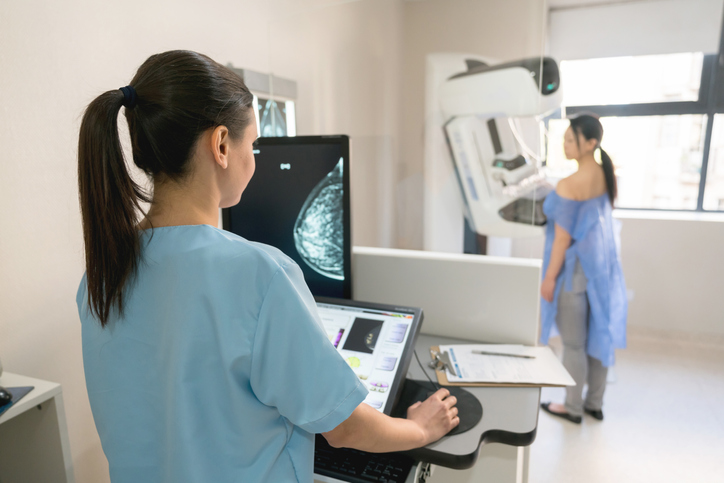
5. Mammography
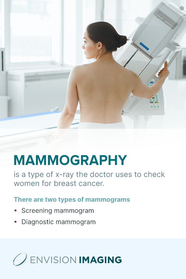
Mammography is a type of x-ray the doctor uses to check women for breast cancer. Mammograms are the pictures it produces. The images might show small tumors you or the doctor can’t feel. Mammograms could also indicate other breast irregularities. There are two types of mammograms.- Screening mammogram: Doctors use this type of mammogram in women with breast cancer, but with no symptoms. The objective is to identify cancer early, when it’s more treatable.
- Diagnostic mammogram: Doctors might perform this test to answer a particular question or to learn more about one of your symptoms.
Benefits of a Screening or Diagnostic Mammogram
Screening mammograms help doctors:- Identify a suspicious area.
- Confirm a lump in your breast.
- Identify the cause of unusual symptoms.
- Take more images of your breast than they’d get with a screening mammogram.
6. Breast Magnetic Resonance Imaging (MRI)
A breast MRI is a diagnostic exam, which your doctor may advise you to get after a mammogram. It captures multiple breast tissue images using magnetic fields. The machine combines the images to generate detailed, computer-generated pictures of the tissue inside your breasts.Benefits of a Breast MRI
A breast MRI can help doctors:- Screen women at high risk for breast cancer.
- Diagnose and assess breast tumors. It does a better job of identifying a small mass in a woman’s breast than an ultrasound or mammogram can, especially if you have non-fatty, dense breast tissue.
- Learn more about cancer identified by feeling a breast but not seen on an ultrasound or mammogram.
- Determine the size of a tumor and how much it spread following a breast cancer diagnosis (staging cancer).
- Monitor the effectiveness of chemotherapy for treating cancer.
- Assess the area where the doctor removed cancerous breast tissue as part of follow-up care.
- Learn if your breast implants ruptured.
7. X-rays
These help your doctor look for cancer in various areas of your body, including your organs like your kidneys and stomach and bones. X-rays are generally quick, painless and without any special preparation required. Contrast studies might need more preparation, and could cause some side effects and discomfort, depending on the type you have. Contrast studies, which are special kinds of x-ray tests, use contrast materials or iodine-based dyes like barium, combined with the x-rays so your organs appear highlighted on the x-ray and the doctor can obtain better images.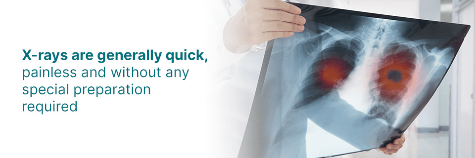
Benefits of X-rays
X-rays don’t cost as much as other scans. Therefore, doctors may use them to obtain information quickly, and before ordering other follow-up tests. An x-ray test is painless and fast. In low doses, the doctor may use them to capture pictures of structures inside your body for detecting and staging a tumor. X-rays produce minimal radiation exposure, and the benefits outweigh the risks, according to research. Doctors can use x-rays in higher doses in radiation therapy to destroy the body’s cancerous cells.8. Nuclear Medicine Scans for Cancer
Nuclear medicine scans generate images based on your body chemistry, instead of on physical forms and shapes, as with other imaging tests. The scans use liquid substances known as radionuclides, radiopharmaceuticals or tracers that release low radiation levels. Body tissues affected by some diseases, like cancer, might absorb more or less of the tracer than normal tissues. Specific cameras pick the radioactivity pattern up to generate images that show the doctor where the tracer travels as well as where it collects. The tumor might show up on the image as a “hot spot” if cancer is present. The “hot spot” is a spot of increased tracer uptake and cell activity. The tumor may also be a “cold spot,” depending on what type of scan the doctor performs. The “cold spot” is the area of less cell activity or decreased uptake. The doctor will determine the type of nuclear scan you’ll receive, depending on which organ they wish to look into. Several common nuclear scans used for cancer include:- PET scans
- Bone scans
- Gallium scans
- Multigated acquisition scans
- Thyroid scans
Benefits of Nuclear Medicine Scans
Nuclear medicine scans help doctors identify tumors, and determine the extent the cancer has spread throughout your body. They also help doctors learn if a treatment is working. They’re relatively painless tests typically performed on an outpatient basis. The tests your doctor recommends might depend on several factors, including:- The location of the tumor and the type it is. Some imaging tests work better than others for different areas of the body.
- Your gender, age and overall health
- Whether or not your doctor requires a biopsy
- Cost
- The balance between any side effects or risks, and the expected benefits
- Your preference
Schedule an Appointment at Envision Imaging for Your Cancer Scan
Book your cancer scan today with Envision Imaging. Our health care specialists are very focused on improving your well-being and health through our hospitality and expertise. We provide affordable, convenient medical services with quick turnaround times, and have committed to ensuring you have as pleasant an imaging case as possible. Consider the following benefits of choosing Envision Imaging for your imaging needs.- Enjoyable and warm patient experience: We treat our patients with the utmost respect, committing to being friendly, helpful and timely. You’ll find our offices are light-filled, elegant and comfortable, and that we show compassion when catering to your needs.
- Fast turnaround rate: We have streamlined our turnaround rates so your doctor can care for your medical needs and get you back to your normal schedule as quickly as possible.
- Most innovative imaging technology: We maintain cutting-edge imaging technology that ensures your procedure results are reliable and accurate.