 Breast cancer is one of the leading cancer diagnoses in women. It is the second-most-common type of cancer in women in the United States — only certain types of skin cancers are more commonly diagnosed. The American Cancer Society reports that one out of every eight women — 13% — will develop breast cancer in her lifetime. Further, one out of every 39 women — about 3% — will die from breast cancer. In 2018, 2 million new cases of breast cancer were diagnosed worldwide.
For women, getting regular mammograms after 40 — or earlier, if particular genetic conditions or health changes raise concerns — is essential for good health. If a woman has breast cancer and her care team can detect it early, it’s much easier to treat. She’ll have a much better chance of surviving to enjoy many more years of life.
When you schedule your exam, you may have a choice between different types of mammograms — two-dimensional (2D) and three-dimensional (3D). Each one takes a particular kind of image that radiologists can use to detect abnormalities early.
This guide will discuss 3D mammography vs. regular mammography to help you determine which method is right for you.
Breast cancer is one of the leading cancer diagnoses in women. It is the second-most-common type of cancer in women in the United States — only certain types of skin cancers are more commonly diagnosed. The American Cancer Society reports that one out of every eight women — 13% — will develop breast cancer in her lifetime. Further, one out of every 39 women — about 3% — will die from breast cancer. In 2018, 2 million new cases of breast cancer were diagnosed worldwide.
For women, getting regular mammograms after 40 — or earlier, if particular genetic conditions or health changes raise concerns — is essential for good health. If a woman has breast cancer and her care team can detect it early, it’s much easier to treat. She’ll have a much better chance of surviving to enjoy many more years of life.
When you schedule your exam, you may have a choice between different types of mammograms — two-dimensional (2D) and three-dimensional (3D). Each one takes a particular kind of image that radiologists can use to detect abnormalities early.
This guide will discuss 3D mammography vs. regular mammography to help you determine which method is right for you.
What Are 2D Mammograms?
A 2D mammogram — also known as conventional digital mammography — takes low-dose x-ray photos of the breast from the front and side perspectives. The photos combine to create a single image of each breast, though sometimes the images show a little overlapping breast tissue. How does a 2D mammogram work? Before imaging, a technologist places one of the patient’s breasts on a special platform in the 2D mammogram machine. A clear plastic plate then gradually presses down on the breast. The plates continue applying pressure while taking 2D x-ray photographs. After getting both front and side images, the technologist repeats the process for the other breast.What Are 3D Mammograms?
In contrast to 2D mammography, 3D mammography — also known as digital breast tomosynthesis — takes multiple x-ray photos from numerous angles around the breast in a continuous arc. A computer imaging program then compiles all the pictures into comprehensive 3D images of each breast. The 3D images show each layer of the breast tissue, one thin slice at a time. Radiologists can pore over the different layers, looking for the microscopic changes that could indicate the presence of cancer.How Are 2D and 3D Mammograms Similar?
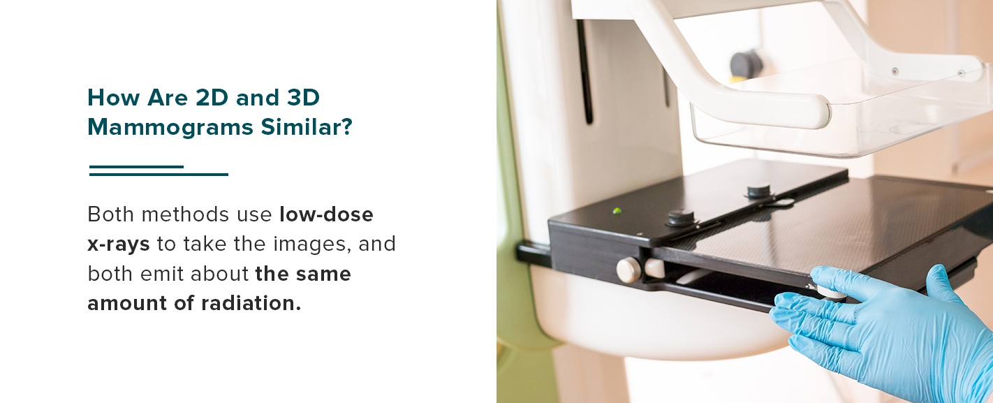 The techniques for 2D and 3D mammograms are similar in their basic mechanisms. Both methods use low-dose x-rays to take the images, and both emit about the same amount of radiation.
The experience of receiving a 2D and 3D mammogram is about the same, though 3D images take a few moments longer to develop. In both cases, a technologist at the clinic or hospital positions the patient’s breast between plastic imaging plates and gradually compresses it for x-ray imaging. The breasts are in the same position for each level of compression during imaging.
The techniques for 2D and 3D mammograms are similar in their basic mechanisms. Both methods use low-dose x-rays to take the images, and both emit about the same amount of radiation.
The experience of receiving a 2D and 3D mammogram is about the same, though 3D images take a few moments longer to develop. In both cases, a technologist at the clinic or hospital positions the patient’s breast between plastic imaging plates and gradually compresses it for x-ray imaging. The breasts are in the same position for each level of compression during imaging.
Schedule a Mammogram With Envision Imaging
How Are 2D and 3D Mammograms Different?
What is the difference between a 2D and 3D mammogram? Images from 2D vs. 3D mammograms differ in their detail, clarity and practicality for certain types of tissue, as well as in their diagnostic applications and insurance coverage. Here is more information on how the two techniques contrast:1. Image Clarity
A 3D mammogram gives the radiologist a clearer picture of the breast tissue and its composition. It brings finer detail into focus and makes tiny structures apparent. Because the tissue is more clearly visible, radiologists can often detect abnormalities they might have missed with a 2D mammogram. One of the limitations of 2D mammography lies in how it compresses the breast tissue and overlaps the x-ray images. The compressing and overlapping can sometimes obscure critical markers in the breast tissue that can indicate cancer. It’s true that 3D mammography also compresses the breast tissue. But because a 3D mammogram enables the radiologist to look at each level of breast tissue individually, slice by slice, the compression does not obscure critical signs. Also, the computer-generated composite ensures there are no overlapping images to interfere with the radiologist’s reading and interpretation.2. Reliability
A 3D mammogram is generally more reliable than a 2D version. In studies, 3D mammography has been shown to have higher cancer detection rates than 2D mammography. A 3D mammogram also makes it less likely for the radiologist to identify a noncancerous structure as cancerous, giving a false positive. This heightened accuracy minimizes unnecessary follow-up imaging — women won’t be called back to the clinic or hospital to have more focused imaging they don’t need — and reduces stress for the misdiagnosed patient.3. Advantages for Denser Tissue
A 3D mammogram is advantageous for all types of patients, though it is optimal for some in particular. Women with denser breast tissue, for instance, may want to choose 3D mammography. Dense breast tissue typically has a higher proportion of glandular structures to fat. In an x-ray image, fatty tissue looks gray, and glandular tissue looks white. However, most cancerous tissue also looks white. With basic 2D mammography, glandular and cancerous structures often look similar. For dense, highly glandular breast tissue, a 3D mammogram is ideal because it can image hidden details of the dense tissue that a basic 2D mammogram might miss. It lets radiologists look at the tissue layer by layer, and it makes distinguishing between glandular and malignant structures more straightforward.
4. Diagnostic Utility
What is the difference between a 3D mammogram and a diagnostic mammogram? A diagnostic mammogram is usually the second type women get if an initial mammogram showed an abnormality. Diagnostic mammograms are more detailed and take longer — the technologist may take more x-ray photographs and zoom in on particular areas of interest as needed. Unlike a 2D mammogram, a 3D mammogram can be useful as a diagnostic tool. If a 2D mammogram reveals an abnormality, the next step is often a biopsy that allows a pathologist to determine whether the tissue is cancerous or benign. Alternatively, your health care providers might use a 3D mammogram for its greater clarity.5. Insurance Coverage
Insurance coverage for 2D and 3D mammograms may differ. Most health insurance plans will cover a standard 2D mammogram, but not all plans will cover a 3D one. Check with your insurance company before scheduling a 3D mammogram to make sure it’s covered under your plan’s terms.Schedule a Mammogram With Envision Imaging Today
Now that you know the difference between a 3D mammogram and a regular mammogram, use that knowledge to keep yourself safe by following the recommended mammogram schedule and choosing the method that will give you the most peace of mind. To protect your health by scheduling a mammogram, contact Envision Imaging. With several locations around the United States, we make it our mission to provide compassionate, trustworthy and effective breast imaging to women across the country. Our comfortable, even spa-like facilities and friendly, knowledgeable technologists will help you have an easy, comfortable scan. You can schedule an appointment with Envision Imaging or the Women’s Center at Colorado Springs Imaging. Contact a location near you to learn more.Call To Schedule Your Appointment
Sources:- https://www.cdc.gov/cancer/breast/statistics/index.htm
- https://www.cancer.org/content/dam/cancer-org/research/cancer-facts-and-statistics/breast-cancer-facts-and-figures/breast-cancer-facts-and-figures-2019-2020.pdf
- https://ww5.komen.org/BreastCancer/Statistics.html
- https://pubs.rsna.org/doi/10.1148/radiol.2019181637
- https://www.envrad.com/services/mammography/
- https://www.envrad.com/locations/