 As a chronic disease, arthritis can take a severe toll on your health, making daily activities more challenging to perform. Some people with arthritis may even experience limited mobility with a decreased range of motion. No matter the severity of the condition, it can complicate everyday life and prevent people from doing what they love to do.
Jump to Sections:
Fortunately, there are many imaging tests available to accurately and efficiently diagnose arthritis. With a prompt diagnosis, you can begin an effective treatment and lifestyle modifications to improve various arthritis symptoms. While there is no cure for arthritis, continuing advancements in technology have vastly improved the effectiveness of different arthritis treatments to minimize painful or uncomfortable symptoms.
As a chronic disease, arthritis can take a severe toll on your health, making daily activities more challenging to perform. Some people with arthritis may even experience limited mobility with a decreased range of motion. No matter the severity of the condition, it can complicate everyday life and prevent people from doing what they love to do.
Jump to Sections:
Fortunately, there are many imaging tests available to accurately and efficiently diagnose arthritis. With a prompt diagnosis, you can begin an effective treatment and lifestyle modifications to improve various arthritis symptoms. While there is no cure for arthritis, continuing advancements in technology have vastly improved the effectiveness of different arthritis treatments to minimize painful or uncomfortable symptoms.
What is arthritis?
Arthritis is when inflammation of a single joint or multiple joints occurs. While there are more than 100 different types of arthritis, two of the most common include osteoarthritis and rheumatoid arthritis. In most cases, arthritis symptoms develop slowly over time and as joints worsen or experience ongoing deterioration. In less common cases, arthritis symptoms may appear suddenly. Arthritis is typically seen in patients over 65, but arthritis can affect patients of all ages. Arthritis is more likely to develop in women and adults who are overweight. While arthritis can cause numerous symptoms, some of the most common symptoms include:- Joint pain
- Stiffness
- Inflammation
- Limited range of motion
- Redness of the skin around affected joint
- Worsening symptoms in the morning
What tests are done to diagnose arthritis?
There are various imaging tests available to detect arthritis that allow a physician to assess the body’s internal structures without an invasive medical procedure. Imaging tests are commonly performed when diagnosing arthritis and monitoring the effectiveness of arthritis treatment. An arthritis imaging test can help detect joints and bone abnormalities that may be a sign of arthritis.X-rays
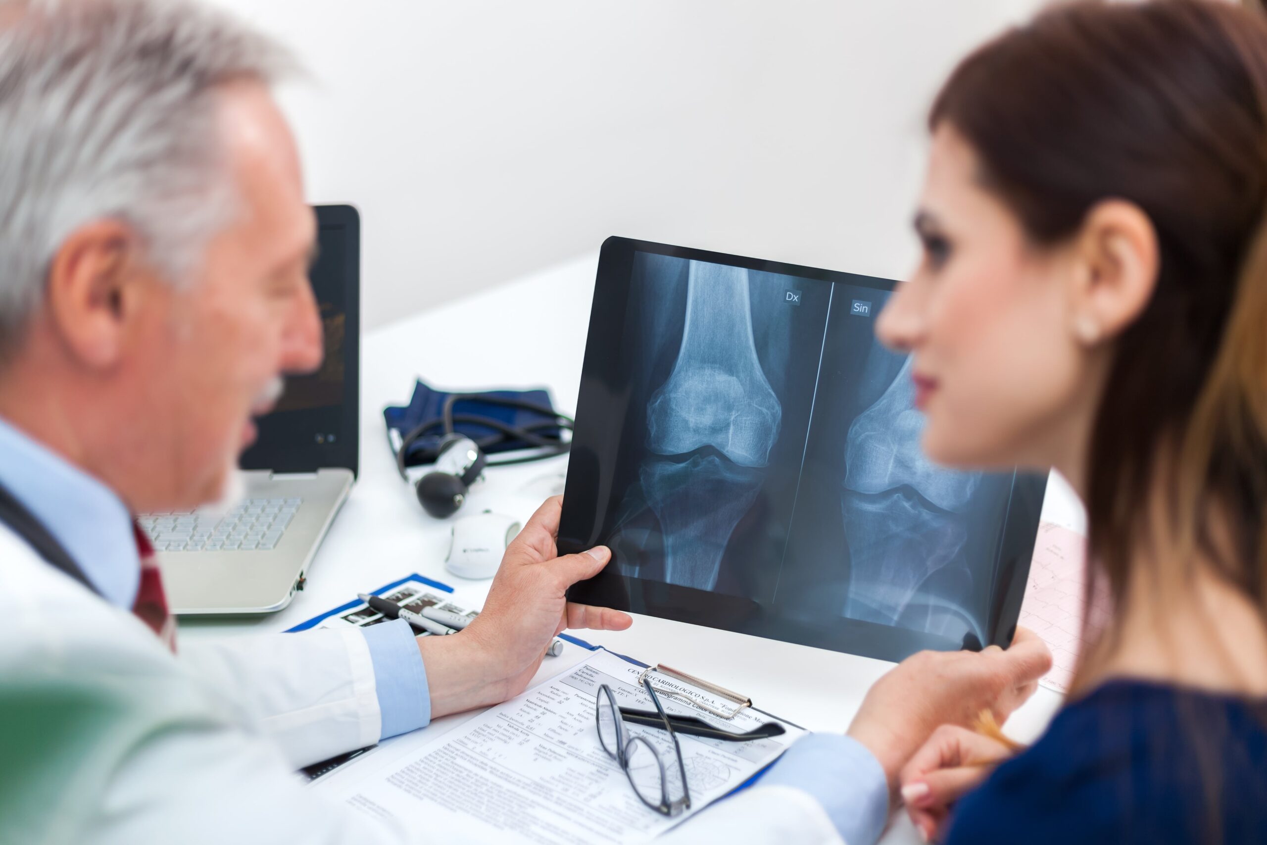
A radiograph, which is more commonly known as an x-ray, uses a safe and low dose of radiation to create images of the body’s internal structures. An x-ray is one of the most common arthritis diagnostic tests used to evaluate, diagnose and monitor degenerative arthritis, inflammatory arthritis and other chronic conditions. An x-ray can show how the bones interact at the joints and are a useful tool to determine the amount of cartilage at the bones’ end. An x-ray can also help detect underlying conditions or deformities that may increase your risk of developing arthritis in the future.Magnetic resonance imaging (MRI)
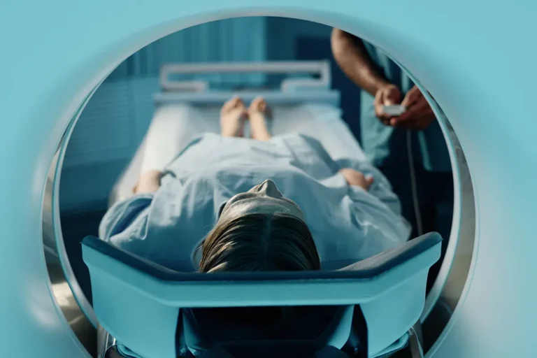
An MRI scan can be a helpful tool when diagnosing arthritis because the scan can create clear images of the body. MRI scans achieve these images by using radio waves, a large magnet and a computer. While it varies from patient to patient, some MRI scans require an injection of a contrast dye or material so doctors can better identify anatomic structures. This arthritis screening test can help diagnose arthritis by evaluating damaged joints, specifically the shoulders, knees and spinal column. In addition to detecting and diagnosing arthritis, an MRI can be a useful tool to track the progression of arthritic joints. MRIs help determine how fast arthritis may be developing or progressing throughout the joints.Computerized axial tomography (CAT)
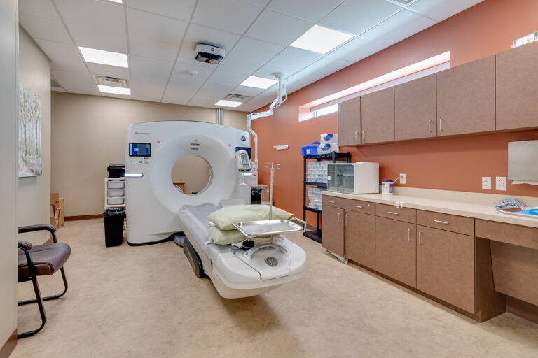
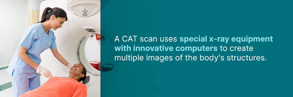 A CAT scan uses special x-ray equipment with innovative computers to create multiple images of the body’s structures. A CAT scan can be a helpful test for different types of arthritis. It can examine the joints deep within the body that traditional x-rays may not be able to examine adequately, including the pelvic region or the spine.
During a CAT scan, the x-ray equipment and computer technology can record clear images of the body, displaying the soft tissues, ligaments and muscles more clearly than a traditional x-ray can. A CAT scan can help gauge the health of the joints and lead to a proper diagnosis of arthritis, helping you receive treatment sooner to minimize discomfort.
A CAT scan uses special x-ray equipment with innovative computers to create multiple images of the body’s structures. A CAT scan can be a helpful test for different types of arthritis. It can examine the joints deep within the body that traditional x-rays may not be able to examine adequately, including the pelvic region or the spine.
During a CAT scan, the x-ray equipment and computer technology can record clear images of the body, displaying the soft tissues, ligaments and muscles more clearly than a traditional x-ray can. A CAT scan can help gauge the health of the joints and lead to a proper diagnosis of arthritis, helping you receive treatment sooner to minimize discomfort.
Bone scan
A bone scan is an innovative radiology procedure that can help examine and detect various bone conditions. A unique aspect of a bone scan is that it can help identify physical and chemical changes in the bone. Bone scans may also be a useful tool to monitor and follow the progression of certain conditions and their treatments. A type of nuclear radiology procedure, a bone scan uses a very small amount of a radioactive substance to aid in examining the bones. The radioactive substance used during a bone scan is commonly referred to as a tracer. This tracer will collect within the bone tissues at spots of physical or chemical abnormalities.Discogram
A discogram, which may be referred to as discography, is a diagnostic imaging test that uses an injection to confirm if there are damaged joints within the spinal column. Discograms are often performed on the lower back to assess the health of the spinal discs. During a discogram, the patient is alert and awake to report if there are any painful or uncomfortable symptoms during the test. A discogram is performed under fluoroscopic guidance to make sure the needle is placed precisely and accurately. A needle is inserted into the disc’s center while a contrast dye is injected. If this injection causes a patient to feel back pain, a physician can determine if the spinal disc may be the source of the pain.Dual-energy x-ray absorptiometry (DEXA)
A DEXA scan may be needed if more specialized imaging tests are required to determine bone health. Physicians may suggest a DEXA scan when diagnosing osteoporosis or arthritis related to the back or feet. During a DEXA scan, a small amount of radiation is used to help determine the density of the bone and overall bone health. When bone density is measured, some patients with advanced osteoarthritis of the spine may appear to have a higher bone density than that measured by other methods. This higher bone density is often the result of bone spurs developing and the discs narrowing, giving a general impression that the bone is thicker than it actually is.World-leading diagnostic imaging at Envision Imaging
Envision Imaging is a leading provider of innovative diagnostic imaging services. We strive to improve lives with exceptional and unmistakable care and service. Envision Imaging and Colorado Springs Imaging offer state-of-the-art radiology services with several locations and is proud to provide a better imaging experience. When you choose Envision Imaging, you can rely on friendly service, knowledgeable team members and world-class radiology services. Learn more about our radiology services and request your radiology screening today. Listed Sources:
- https://www.cdc.gov/arthritis/basics/types.html
- https://www.envrad.com/services/x-ray/
- https://www.envrad.com/services/mri-scans/
- https://www.envrad.com/services/ct-scans/
- https://www.envrad.com/services/bone-density/
- https://www.envrad.com/services/dexa/
- https://www.envrad.com/about-us/
- https://www.envrad.com/locations/
- https://www.envrad.com/services/