Whether you’ve received a melanoma diagnosis or noticed abnormal skin, you’ll need tests to gather more information. Melanoma is one of the most common forms of cancer, with tens of thousands of patients receiving a diagnosis each year.
Imaging technologies are drastically affecting melanoma diagnoses and treatments to help patients get the care they need faster and more efficiently. Imaging tests can identify the disease and monitor whether it’s spreading or recurring after treatment. Continue reading to learn how to test for skin cancer with imaging tests and what to expect.
How do doctors test for melanoma?
Doctors rely on a variety of tests to determine whether a patient has melanoma. Blood tests, physical exams, biopsies and imaging tests provide in-depth information about the condition and how to treat it. Your medical history can also detail information about your condition. For example, a history of sunburns and tanning and a family history of skin cancer can give insights into the signs and symptoms you’re experiencing.
How to test for melanoma using imaging tests
Imaging tests can be helpful indicators of melanoma because they provide pictures of the inside of your body through radioactive substances, x-rays or magnetic fields. These tests allow doctors to determine whether the melanoma has spread to other organs or lymph nodes.
Imaging tests can help your provider evaluate the effectiveness of treatment and monitor whether cancer is recurring after treatment. Many early-stage melanoma patients do not need imaging tests because the cancer is unlikely to have spread. Below, you can find more information about imaging tests for detecting melanoma and what to expect from each.
Chest x-ray
An x-ray creates images of your organs and bones using small radiation doses. The x-ray machine will emit a safe radiation level that passes through your body to record images on a specialized plate. Chest x-rays detail whether the melanoma has spread throughout your body. You can use this exam specifically to determine whether there have been any changes in your lungs. This test can also identify whether you have enlarged lymph nodes in your chest.
A technologist will typically position your body to ensure they can gather clear pictures. You may need to hold your breath and remain still to avoid blurring the image. This imaging test typically lasts for a few minutes and does not cause discomfort or pain.
Ultrasound
Ultrasound tests form images of your body using sound waves. Ultrasound scanners have a microphone that emits sound waves, which bounce around your body for the microphone to pick up again. The results show up on a computer and form a complete picture.
This test involves a specialist moving a probe with lubricating gel across your skin. Although this test is not painful, some patients can experience discomfort as the probe moves across their skin. If this occurs, inform the specialist as soon as possible. Your exam will end if your test results indicate a normal-looking lymph node.
However, if you have an abnormal looking-lymph node, your provider can use your ultrasound to guide biopsy needles into the lymph node to gather more information. To do so, they will guide a fine needle through your skin and use a syringe to draw fluid and cells. This process will allow your doctor to gather samples of tissue or cells, which a specialist can evaluate under a microscope to determine whether cancer cells are present.
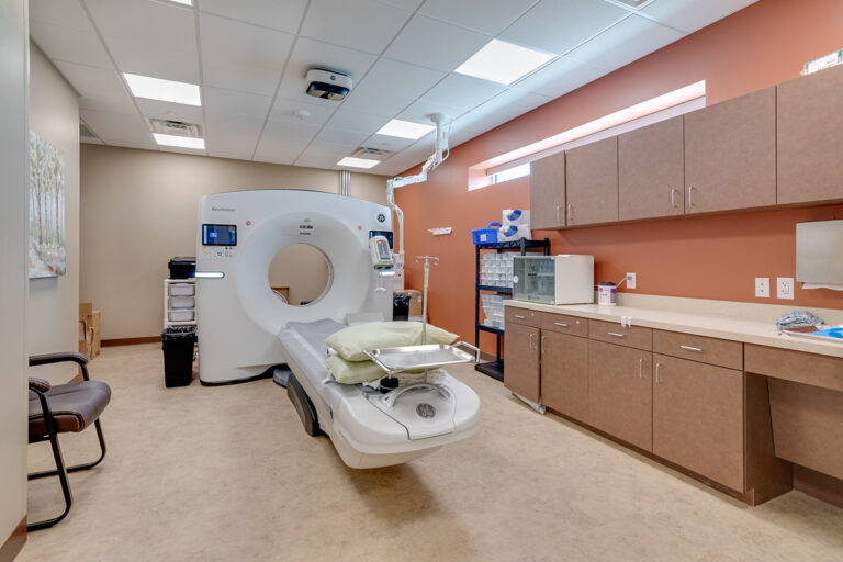
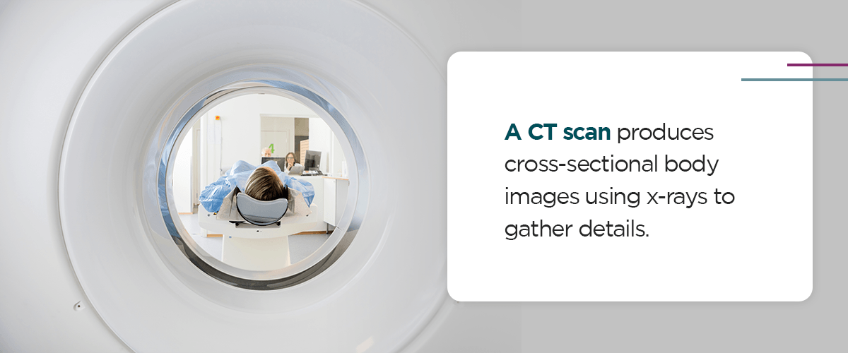
CT scan
A CT scan produces cross-sectional body images using x-rays to gather details. The machine will capture images from different angles during the test to create a 3D picture on a computer. CT scans can detail soft tissue, like internal organs, making it a more comprehensive examination than traditional x-rays.
This exam can depict whether you have enlarged lymph nodes or if your liver, lungs or other organs have suspicious spots, which could be due to the melanoma spreading. CT scans can also guide your provider to insert a biopsy needle into a suspicious area.
Before the exam, you may need to drink or receive an injection of contrast dye. This material allows the results to show more clearly on the computer. You may also need to have a blood test prior to the CT scan to ensure your kidneys can flush the dye after the scan is complete.
This exam typically takes a few minutes and does not cause pain. However, you may feel a hot sensation or get uncomfortable while remaining still. If you experience claustrophobia, speak with your provider before the exam to ensure a more pleasant experience.
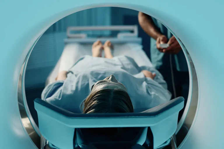
MRI scan
Magnetic resonance imaging (MRI) scans require magnets and radio waves to complete detailed images of your body. This scan will create a cross-sectional image that enables your provider to evaluate all areas of your body and examine soft tissue. These exams are especially helpful when you need to evaluate your brain or spinal cord, and they can detect whether the melanoma has spread to other areas.
These scans have a similar process to CT scans — you will need a contrast medium before entering the MRI machine. Though painless, MRI scans require strong magnetism, which can affect any metal in your body. You should inform your provider if you have a pacemaker, surgical plates, pins or clips, cochlear implants, or metal fragments such as dental fillings or possible fragments from an injury.
PET scan
Position emission tomography (PET) scans are beneficial for patients with advanced stages of melanoma. During this scan, you will receive a slight radioactive injection so that a special camera can collect radioactivity in your body. The injection will typically be a form of sugar. This allows your provider to determine where your body uses the most sugar to find active cells, like cancer cells.
Similar to an MRI or CT scan, you will slide into the scanner and remain still while the machine collects images. It can take a half hour or longer to complete this exam, and the procedure is painless. Some healthcare facilities use a PET-CT scan or PET-MRI machine to perform two tests simultaneously. This enables your provider to better assess radioactive areas with a more detailed appearance.
Test for melanoma with Envision Imaging
At Envision Imaging, we aim to provide the most convenient and compassionate care in your area, so you can find accurate imaging anytime you need it. Our warm hospitality and strong patient focus empower us to help each patient find the care they deserve while putting each person at ease.
With us, you can find comprehensive care and precise imaging services. We are revolutionizing the radiology field with spectacular service and unmistakable quality. Browse our locations to request your next diagnostic imaging appointment. Our world-class facilities, compassionate care and accurate results ensure a pleasant and reliable experience.

 This exam can depict whether you have enlarged lymph nodes or if your liver, lungs or other organs have suspicious spots, which could be due to the melanoma spreading. CT scans can also guide your provider to insert a biopsy needle into a suspicious area.
Before the exam, you may need to drink or receive an injection of contrast dye. This material allows the results to show more clearly on the computer. You may also need to have a blood test prior to the CT scan to ensure your kidneys can flush the dye after the scan is complete.
This exam typically takes a few minutes and does not cause pain. However, you may feel a hot sensation or get uncomfortable while remaining still. If you experience claustrophobia, speak with your provider before the exam to ensure a more pleasant experience.
This exam can depict whether you have enlarged lymph nodes or if your liver, lungs or other organs have suspicious spots, which could be due to the melanoma spreading. CT scans can also guide your provider to insert a biopsy needle into a suspicious area.
Before the exam, you may need to drink or receive an injection of contrast dye. This material allows the results to show more clearly on the computer. You may also need to have a blood test prior to the CT scan to ensure your kidneys can flush the dye after the scan is complete.
This exam typically takes a few minutes and does not cause pain. However, you may feel a hot sensation or get uncomfortable while remaining still. If you experience claustrophobia, speak with your provider before the exam to ensure a more pleasant experience.