If you have a condition that affects your joints, bones or muscles, you are most likely dealing with a musculoskeletal disorder. Musculoskeletal disorders are relatively common, and the risk of developing one increases as you get older. Other factors that can increase your risk of developing a musculoskeletal disorder include the type of activities you participate in, your overall activity level and your family history.
Treating a musculoskeletal disorder depends on getting the diagnosis right. Fortunately, there are many imaging options available to aid medical providers in musculoskeletal diagnosis. Along with imaging, your medical provider might order blood tests and conduct a physical exam to pinpoint the root cause and give you the best treatment possible.
Imaging Tests Used for Diagnosing Muscle Disorders
Your medical provider might order one or more imaging tests for a closer look at your muscles, joints and bones. The type of scan for muscle damage they order will depend largely on what they think the issue is. Here are more details on a few of the imaging tests used to diagnose musculoskeletal disorders.
CT Scan
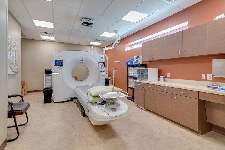
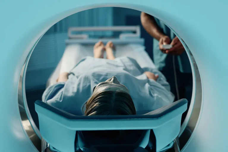
Doctors often use a computed tomography scan, aka a CT scan or CAT scan, to diagnose problems with the bones or muscles. A CT scan takes x-ray pictures from various angles. It provides a more in-depth look at the interior of the body than imaging options such as an x-ray.
During a CT scan, you lie on a table and pass through a large, ring-shaped scanner. Straps and pillows on the table might hold you in place. While you pass through the scanner, it takes images of your body. The technologist operating the scanner is usually in a separate room, just off to the side. They gather the CT images and assemble them into three-dimensional pictures that a medical provider can review to determine what’s going on.
Usually, a CT scan is best to capture pictures of your bones.
DEXA Scan
A DEXA scan measures the density and mass of structures inside the body. A common use for this type of imaging is to measure bone mass, such as in patients with osteoporosis. Like a CT scan, a DEXA scan uses x-ray imaging. It’s an acronym for “dual-energy x-ray absorptiometry.”
During a DEXA scan, you lie flat on a table. The scanner passes over you, from above, using two different types of x-ray beams to take pictures. The entire imaging process typically takes up to 30 minutes.
While health providers commonly use a DEXA scan to measure bone mass, it can also assess fat and muscle mass.
X-Ray
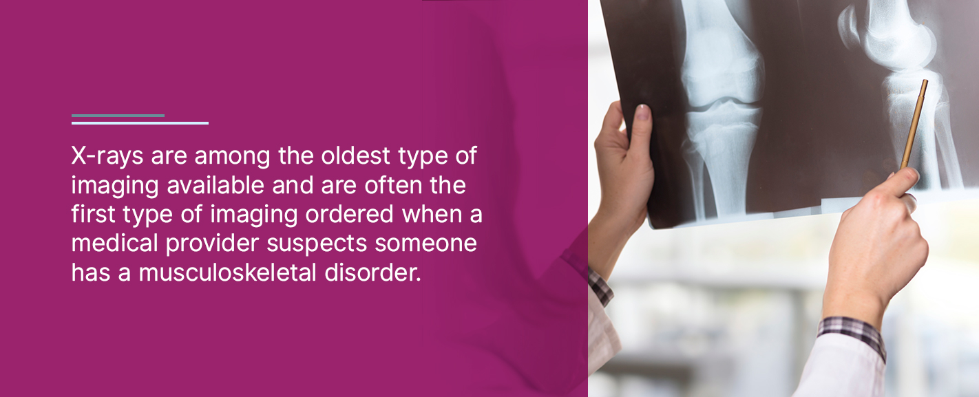
X-rays are among the oldest type of imaging available and are often the first type of imaging ordered when a medical provider suspects someone has a musculoskeletal disorder. The imaging uses electromagnetic waves to produce an image. How organs and bones appear on the x-ray image depends on how well the magnetic waves pass through them. Bones, which are primarily calcium, absorb the energy waves well, so they usually show up as white on the imaging. If there’s a break in the bone, it shows up black, as the waves of energy can pass through it.
Do x-rays show muscles? Though a health provider might order an x-ray to determine a muscular disorder, muscles and other types of soft tissues usually don’t show up on x-rays. They are likely to appear grayish, as the magnetic waves pass through them relatively easily.
If a provider wants to see evidence of damage to a joint, they might order a stress x-ray, taken while moving or positioning a joint at several different angles.
MRI
Magnetic resonance imaging, aka MRI, uses radio waves and magnetic fields to capture pictures inside the body. During an MRI, a patient lies very still and travels through a long, tube-shaped machine. The imaging produces three-dimensional pictures that look like cross-sections of the inside of the body. Since the imaging uses magnets, it’s not ideal for people with metal plates, implants or pacemakers.
Since some patients find the traditional MRI tube confining or anxiety-inducing, open MRI scanners are also available. These scanners are open on three sides, creating a more comfortable experience.
Does an MRI show muscle damage? Unlike x-ray imaging, MRI imaging is excellent at capturing images of the soft tissues of the body, including the muscles. It can show muscle damage due to a musculoskeletal disorder. MRI imaging also captures joint damage well, such as torn cartilage or ligaments.
Arthrogram
An arthrogram is a type of imaging that takes a picture of the inside of the joint. The imaging can be a type of x-ray, CT or MRI, depending on the issue. During an arthrogram, contrast dye gets injected into the joint. The technologist moves the joint into various positions while taking images.
Health professionals usually perform an arthrogram for patients with unexplained joint issues, such as pain with movement or the loss of motion.
One potential concern associated with arthrography is that some people might be allergic to the dye used to improve the images. If you’ve ever had an allergic reaction to contrast dye or know that you have an allergy to the ingredients commonly found in the dye, let your medical provider and the technologist known before the imaging begins. They might choose a different type of imaging test instead.
Ultrasound
An ultrasound, or sonography, uses soundwaves to take pictures of the inside of the body. Ultrasounds are excellent at capturing images of the soft tissues, including muscles and ligaments. Though you might associate ultrasound imaging with pregnancy, it can also diagnose musculoskeletal disorders. The imaging is particularly effective at picking up signs of swelling and inflammation near joints and muscles.
Schedule an Imaging Appointment at Envision Imaging Today
Which type of imaging will help you get the most accurate diagnosis? Your medical provider is likely to start with an x-ray, then order additional tests if they need more guidance or want to take a closer look. The type of test they order might also depend on what they think might be the issue. X-ray imaging is often appropriate for detecting issues with the bones, while an MRI might be more appropriate if a doctor suspects any issues with your body’s soft tissues.
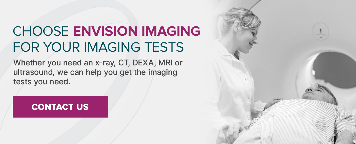
Whether you need an x-ray, CT, DEXA, MRI or ultrasound, Envision Radiology has locations across Colorado, Louisiana, Oklahoma and Texas to help you get the imaging tests you need. Find the location nearest you to schedule an imaging appointment today.

 If you have a condition that affects your joints, bones or muscles, you are most likely dealing with a musculoskeletal disorder. Musculoskeletal disorders are relatively common, and the risk of developing one increases as you get older. Other factors that can increase your risk of developing a musculoskeletal disorder include the type of activities you participate in, your overall activity level and your family history.
Treating a musculoskeletal disorder depends on getting the diagnosis right. Fortunately, there are many imaging options available to aid medical providers in musculoskeletal diagnosis. Along with imaging, your medical provider might order blood tests and conduct a physical exam to pinpoint the root cause and give you the best treatment possible.
If you have a condition that affects your joints, bones or muscles, you are most likely dealing with a musculoskeletal disorder. Musculoskeletal disorders are relatively common, and the risk of developing one increases as you get older. Other factors that can increase your risk of developing a musculoskeletal disorder include the type of activities you participate in, your overall activity level and your family history.
Treating a musculoskeletal disorder depends on getting the diagnosis right. Fortunately, there are many imaging options available to aid medical providers in musculoskeletal diagnosis. Along with imaging, your medical provider might order blood tests and conduct a physical exam to pinpoint the root cause and give you the best treatment possible.
 X-rays are among the oldest type of imaging available and are often the first type of imaging ordered when a medical provider suspects someone has a musculoskeletal disorder. The imaging uses electromagnetic waves to produce an image. How organs and bones appear on the x-ray image depends on how well the magnetic waves pass through them. Bones, which are primarily calcium, absorb the energy waves well, so they usually show up as white on the imaging. If there’s a break in the bone, it shows up black, as the waves of energy can pass through it.
Do x-rays show muscles? Though a health provider might order an x-ray to determine a muscular disorder, muscles and other types of soft tissues usually don’t show up on x-rays. They are likely to appear grayish, as the magnetic waves pass through them relatively easily.
If a provider wants to see evidence of damage to a joint, they might order a stress x-ray, taken while moving or positioning a joint at several different angles.
X-rays are among the oldest type of imaging available and are often the first type of imaging ordered when a medical provider suspects someone has a musculoskeletal disorder. The imaging uses electromagnetic waves to produce an image. How organs and bones appear on the x-ray image depends on how well the magnetic waves pass through them. Bones, which are primarily calcium, absorb the energy waves well, so they usually show up as white on the imaging. If there’s a break in the bone, it shows up black, as the waves of energy can pass through it.
Do x-rays show muscles? Though a health provider might order an x-ray to determine a muscular disorder, muscles and other types of soft tissues usually don’t show up on x-rays. They are likely to appear grayish, as the magnetic waves pass through them relatively easily.
If a provider wants to see evidence of damage to a joint, they might order a stress x-ray, taken while moving or positioning a joint at several different angles.
 Which type of imaging will help you get the most accurate diagnosis? Your medical provider is likely to start with an x-ray, then order additional tests if they need more guidance or want to take a closer look. The type of test they order might also depend on what they think might be the issue. X-ray imaging is often appropriate for detecting issues with the bones, while an MRI might be more appropriate if a doctor suspects any issues with your body’s soft tissues.
Whether you need an x-ray, CT, DEXA, MRI or ultrasound, Envision Radiology has locations across Colorado, Louisiana, Oklahoma and Texas to help you get the imaging tests you need. Find the location nearest you to schedule an imaging appointment today.
Which type of imaging will help you get the most accurate diagnosis? Your medical provider is likely to start with an x-ray, then order additional tests if they need more guidance or want to take a closer look. The type of test they order might also depend on what they think might be the issue. X-ray imaging is often appropriate for detecting issues with the bones, while an MRI might be more appropriate if a doctor suspects any issues with your body’s soft tissues.
Whether you need an x-ray, CT, DEXA, MRI or ultrasound, Envision Radiology has locations across Colorado, Louisiana, Oklahoma and Texas to help you get the imaging tests you need. Find the location nearest you to schedule an imaging appointment today.