 Blood clotting in the body is often beneficial. It helps staunch wounds, prevent infections and begin the healing process after an injury. But when blood clots form in the blood vessels, they pose grave health risks.
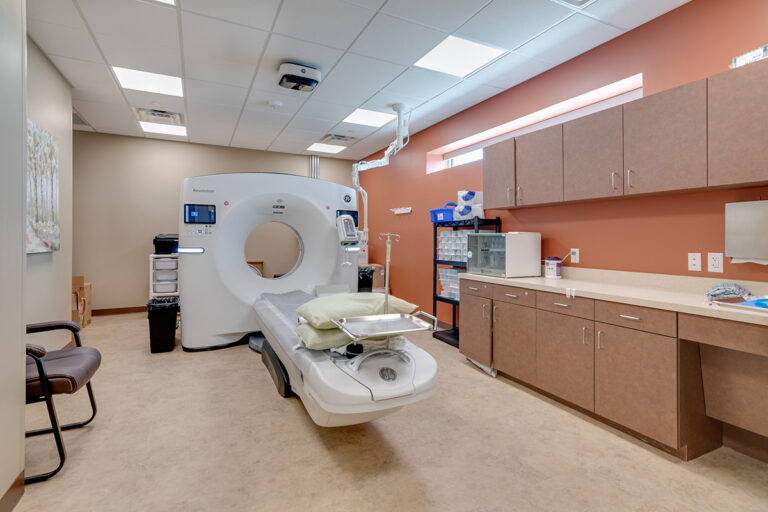
Fortunately, computed tomography (CT) scans provide valuable detection and diagnostic information. How does a CT scan show blood clots? In this article, we discuss how health care professionals scan for blood clots and how these CT techniques work.
Blood clotting in the body is often beneficial. It helps staunch wounds, prevent infections and begin the healing process after an injury. But when blood clots form in the blood vessels, they pose grave health risks.
Fortunately, computed tomography (CT) scans provide valuable detection and diagnostic information. How does a CT scan show blood clots? In this article, we discuss how health care professionals scan for blood clots and how these CT techniques work.
What Are Blood Clots?
Blood clots are gel-like or semisolid clumps in the bloodstream. Under normal, healthy conditions, blood flows freely through the arteries and veins of the body. Sometimes, though, the blood begins to congeal in one place and adhere to vein or artery walls. Depending on their location and severity, blood clots may be exceptionally dangerous, even life-threatening. Blood clots typically come in one of two different types — thromboses or embolisms.- Thrombosis: A thrombosis is a blood clot that remains stationary in the bloodstream. It impedes blood flow, forcing the blood to squeeze through the small space next to the clot.
- Embolism: An embolism is a blood clot that breaks free from its original location and travels elsewhere. When these clots end up in the lungs, for instance, they are known as pulmonary embolisms.
Arterial Clots
Just as doctors can classify blood clots by how they behave in the bloodstream, they can also classify blood clots by where they form. Arterial clots form in the body’s arteries — the blood vessels that carry blood away from the heart and toward the body’s other organs and tissues. Arterial clots cause immediate symptoms. Arterial blood carries vital oxygen from the heart to other areas of the body. If a blood clot interferes with that oxygen transport, the person may experience a severe medical event such as a stroke, heart attack, sudden paralysis or extreme pain.Venous Clots
Venous clots form in the veins — the smaller blood vessels that carry oxygen-depleted blood from the organs and tissues back to the heart. Venous blood clots generally form slowly, and because they do not interfere with the body’s oxygen supply, they may be symptomless at first. As time passes, though, the person may begin to experience symptoms, or previously mild symptoms may become much more noticeable.What Causes Blood Clots?
 Numerous factors — some hereditary, some lifestyle-related — can make blood clot formation more likely:
- Advanced age — especially 65 or older
- Bed rest or other long sedentary periods, such as during travel
- Cancer
- Family history of blood clots
- Medications like hormonal birth control
- Obesity
- Pregnancy
- Smoking
Where Do Blood Clots Form?
Blood clots commonly form in several areas of the body:- Abdomen: Blood clots that form in the abdomen can cause severe pain and gastrointestinal symptoms like diarrhea, nausea, vomiting and blood in the stool.
- Arms and legs: Blood clots that form in the arms and legs can cause bruises, cramps, pain, swelling and excessively warm skin. Deep vein thrombosis (DVT) occurs when stationary clots form in the larger, deeper veins, and they tend to fly under the radar —and according to the U.S. Centers for Disease Control and Prevention (CDC), about 50% of people with deep vein thrombosis experience no symptoms.
- Brain: A blood clot in the brain can often lead to a stroke. These clots cause neurological symptoms such as vision impairment, slurred speech, bodily weakness and seizures.
- Heart: A blood clot in the heart can often lead to a heart attack. These clots cause chest pain that radiates into the left arm, as well as sweating and labored breathing. Clots in the heart also often travel on to the brain or lungs.
- Lungs: In the lungs, blood clots can cause coughing, chest pain and difficulty breathing. Less commonly, they also cause dizziness, irregular heartbeat and pulse, swelling of the extremities, skin discoloration and excessive sweating.
How CT Scans Detect and Diagnose Blood Clots
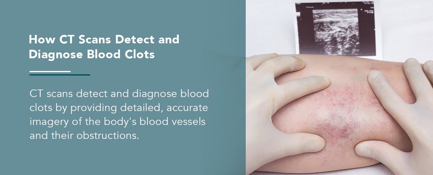 CT scans detect and diagnose blood clots by providing detailed, accurate imagery of the body’s blood vessels and their obstructions. Doctors generally use two CT scan techniques for blood clot detection and diagnosis — CT venography and CT pulmonary angiography.
CT Venography
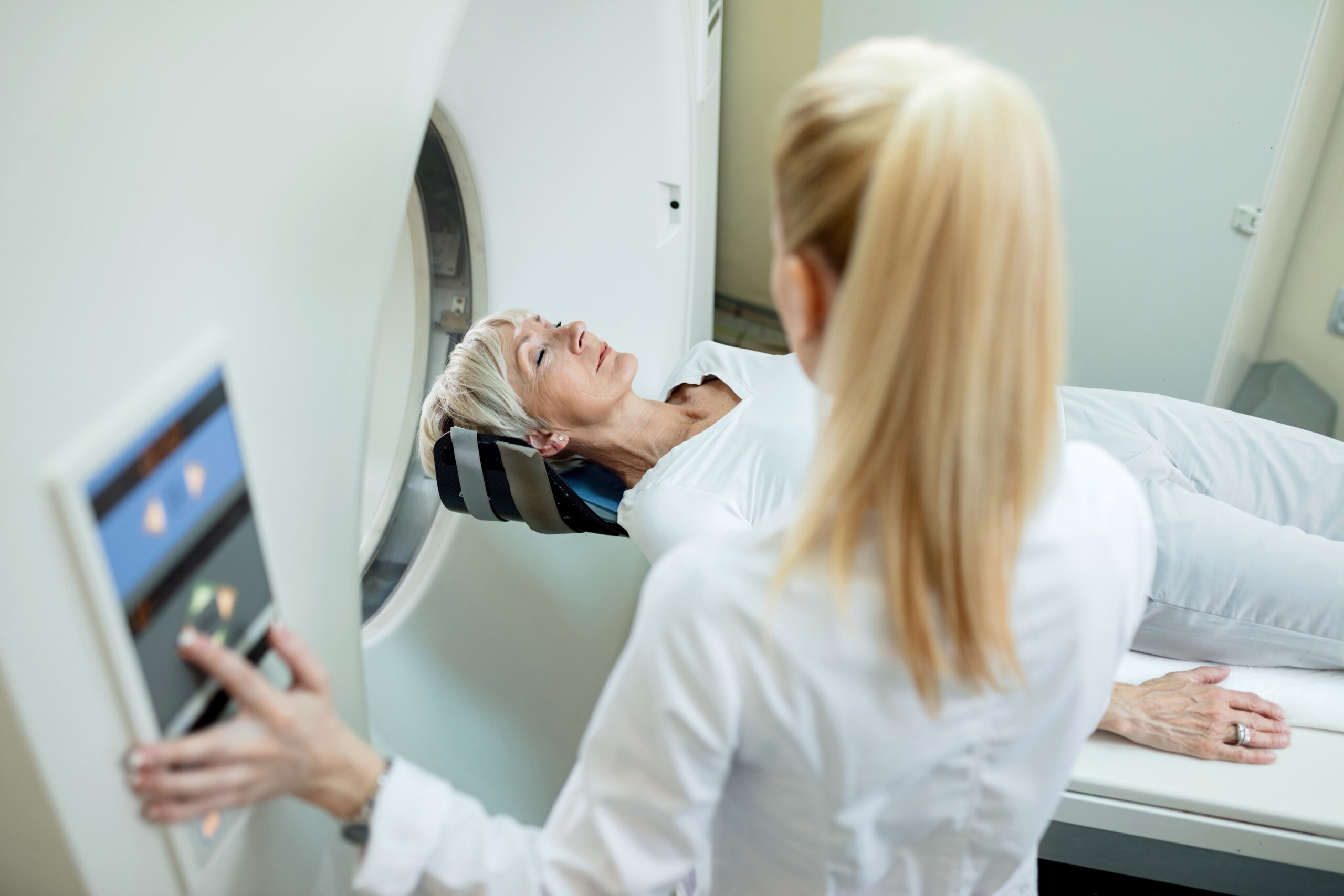
CT venography (CTV) creates maps of the veins, often the veins in the legs, by using contrast dye, advanced x-ray technology and computer imaging. To start, a technologist injects an iodine-based contrast dye into one of the patient’s veins, usually through an intravenous (IV) line. The contrast dye spreads throughout the nearby bloodstream and shows up clearly on x-rays. Veins do not otherwise show up well on x-rays, so the contrast dye is necessary for an accurate image. Next, a medical professional takes radiographs or x-rays of the area where doctors suspect blood clots. Unlike traditional radiography, which uses a single x-ray source that bounces off a detector plate to create an image, CT scanning uses a chamber with multiple x-ray sources and detectors that spin around the body at tremendous velocity. Then a computer processes the images from the scanner to create detailed, clear images. On the CT scan, blood clots and blood vessels show up clearly in a whitish color because of the contrast dye. A radiologist then analyzes the images and sends a report to the referring physician.CT Angiography
CT angiography (CTA) maps the arteries, often the arteries that carry blood from the heart to the lungs. When applied to the lungs, this technique is called CT pulmonary angiography. Like CT venography, CT angiography uses an injectable contrast dye to make the arteries to show up well for imaging. Then a CT scanner takes a series of radiographs and sends them for computer processing and a radiologist’s analysis of any observed blood clots.Additional Imaging Tests That Can Aid in Diagnosing Blood Clots
Beyond CT imaging, doctors can use a few different tests to help detect and diagnose blood clots:- Venous ultrasound: Venous ultrasound is similar to CT venography but is less invasive. It uses sound waves to create a map of the veins without requiring injectable dye or radiation exposure. It is not always as informative as CT venography, though, so if venous ultrasound results are inconclusive, venography is often the next step.
- MR angiography: Magnetic resonance angiography (MRA) maps the arteries using magnetic resonance imaging (MRI). It uses a magnetic field, radio waves and computer imaging. The magnetic field and radio waves safely realign the body’s hydrogen atoms, and when the hydrogen atoms regain their customary alignment, they emit specific amounts of energy. The scanner captures that energy and uses it to create an image of the arteries and any blood clots they contain.
- Ventilation-perfusion scan: A ventilation-perfusion (V/Q) scan uses special radioactive dye to examine ventilation and perfusion — that is, airflow and blood flow — to the lungs and check for blockages.
- Echocardiography: Health care professionals frequently use echocardiography when patients are already exhibiting signs of a stroke. To reach the cerebral arteries, an embolism typically either forms in the heart or travels through the heart. An echocardiogram uses ultrasound imaging to inspect the blood vessels of the heart for confirmation.
Schedule an Appointment for Diagnostic Imaging With Envision Imaging and Colorado Springs Imaging
If you or your medical provider suspects clots in your blood vessels, the best thing you can do is to make an appointment for diagnostic imaging right away. Make Envision Imaging and Colorado Springs Imaging your trusted source to scan for blood clots. We provide both CTA services and MRA scans, and we also offer responsive and flexible scheduling, superior image quality and fast turnaround times for results. You’ll experience a compassionate, convenient process and get your results quickly so you can move on to receiving the treatment you need. Find a Colorado Springs Imaging (CO) or Envision Imaging (TX / LA / OK) location near you to learn more.Call To Schedule Your Appointment
Sources:- https://www.healthline.com/health/how-to-tell-if-you-have-a-blood-clot
- https://www.cdc.gov/ncbddd/dvt/facts.html
- https://www.radiologyinfo.org/en/info.cfm?pg=venography
- https://www.radiologyinfo.org/en/info.cfm?pg=angioct
- https://www.radiologyinfo.org/en/info.cfm?pg=venousus
- https://www.radiologyinfo.org/en/info.cfm?pg=angiomr
- https://www.insideradiology.com.au/vq-scan
- https://www.verywellhealth.com/how-blood-clots-are-diagnosed-4163662
- https://www.envrad.com/services/ct-scans/
- https://www.envrad.com/services/mra-scans/
- https://www.envrad.com/locations/