Women’s imaging is image tests specifically for women’s health. While this may seem obvious, there is far more to this sector of radiology than that. Because women have different health concerns than men, they need image tests designed especially for them. From helping women catch diseases before they develop into larger issues to monitoring pregnancy, women’s imaging delivers this specialized care. This article will explore the field of women’s imaging, along with the main types of women’s imaging and where to find a dependable women’s imaging specialist.
What Is Women’s Imaging
Women’s imaging covers several diagnostic imaging procedures that specifically apply to women and determine diseases that are most common in females, such as gynecological complications or breast cancer. This women-focused branch of radiology is largely concerned with achieving an early diagnosis so a disease may be treated as quickly and effectively as possible.
These are some of the most notable benefits of diagnostic imaging for women:
Diagnostic imaging tests, like digital screening, can provide patients with immediate results.
Some women’s imaging tests, like mammograms, can detect cancer years in advance of the disease’s first visible signs.
For recovering cancer patients, women’s imaging tests can monitor the after-effects of treatment and alert them of a recurrence.
Imaging tests, like digital mammograms and breast ultrasounds, can help women who have dense breast tissue be aware of whether they have an underlying health issue they need to address.
Certain tests can be used to screen women for diseases prevalent in females, especially those who are more advanced in age.
Although imaging tests do not help prevent or treat a disease, they can increase a patient’s chances of survival and recovery by catching the disease before it can turn into a larger medical issue.
By providing more information about the patient’s condition, women’s imaging tests can help them and their doctors make decisions regarding the course of their treatment going forward.
Types of Women’s Imaging Tests
The array of women’s imaging services includes the following types of tests:
1. Mammogram
A mammogram is a common imaging test for women that takes an x-ray picture of the breast. Doctors look at the mammogram picture to detect early signs of breast cancer, such as lumps that are not big enough for the patient to feel or abnormal changes in the breast tissue. For this reason, regular mammograms are crucial to help catch breast cancer early so it can be addressed as soon as possible. By receiving routine mammograms, women can find breast cancer as much as three years before it can be felt, in some cases.
Digital mammogram imaging is the most reliable method for locating and diagnosing cancer nodules because older mammogram systems tend to produce a less clear picture. Within digital mammograms, there are two types — screening mammography and diagnostic mammography. These are the main differences between the two:
Screening mammography: This type of mammogram is performed annually to find unsuspected breast cancer in women, even if no other symptoms or signs are present. This is an important component of a regular preventive health care screening program.
Diagnostic mammography: Women may receive a diagnostic mammogram if a concerning area is found during the screening exam. For abnormal clinical findings or breast disease symptoms like a palpable lump, skin dimpling or nipple discharge, a doctor may recommend a diagnostic mammogram.
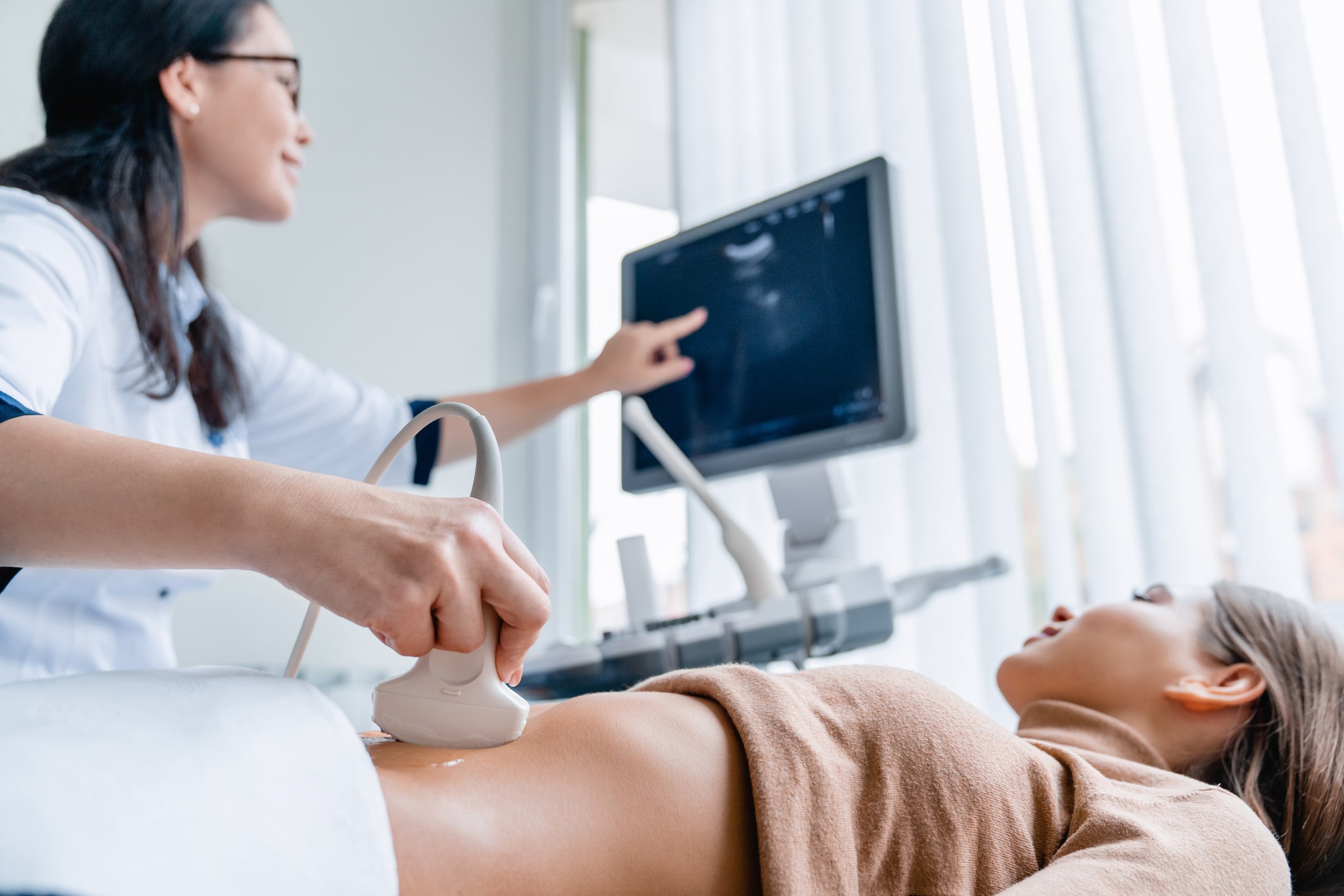
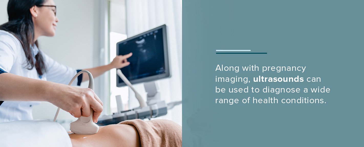
An ultrasound uses high-frequency sound waves to deliver real-time images of the inside of a patient’s body, including blood flow and the structure of internal organs. By using sound waves that can travel through soft tissue and fluids but bounce back upon hitting denser surfaces, an ultrasound can produce an accurate picture. Because the test does not use radiation, it is a safe technique to use during pregnancy.
Along with pregnancy imaging, ultrasounds can be used to diagnose a wide range of health conditions. For example, breast ultrasounds are used to detect breast tissue abnormalities or explore potential abnormalities found in a mammogram. With an ultrasound, a doctor may be able to determine whether a breast lump is a benign cyst or potentially a cancerous tumor. In addition, a breast ultrasound can help medical professionals target the correct area for a breast biopsy.
3. Breast Biopsy
When a lump, mass or other abnormality is found on the breast, a breast biopsy is often done to determine whether the mass is cancerous or benign. During the breast biopsy, a tissue sample is taken and sent to a lab for further testing. Breast biopsies are typically guided by an imaging test, such as an MRI-guided breast biopsy or an ultrasound-guided breast biopsy.
4. Breast MRI
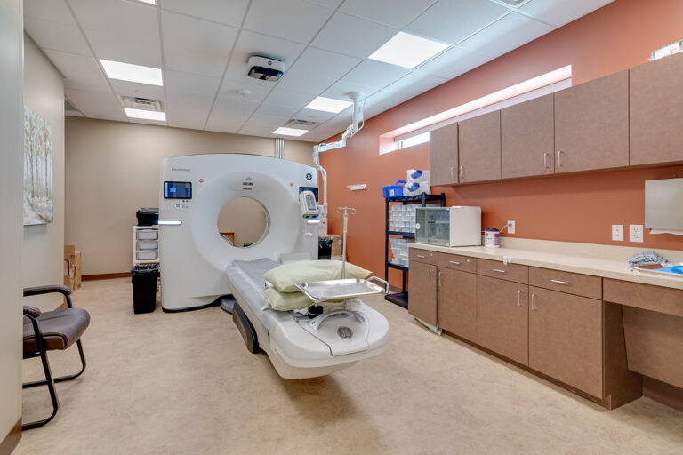
A breast MRI can provide doctors with even more information than a mammogram or ultrasound. If a medical professional needs more information than a clinical breast exam, mammogram or ultrasound can give, they will often recommend a breast MRI because the procedure uses radiation to produce more detailed images.
For certain patients, such as those with a very high risk of breast cancer, a breast MRI might be used as a screening tool like a mammogram would. Although a breast MRI should not serve as a replacement for a mammogram or ultrasound, it can be helpful as an additional tool for detecting breast abnormalities and identifying breast cancer quickly.
5. Bone Density Scan
Also called bone mineral density testing, a bone density scan can be used as an indicator of osteoporosis. By using x-rays to measure the amount of mineral material per square centimeter, this imaging test can detect the density of a patient’s bones. This can help doctors determine the chances of fracture for women with a lower bone density and lack of minerals.
Because women typically have thinner, smaller bones than men, as well as a sharp decline in estrogen — a hormone that protects bones — with age, it is important for women who have gone through menopause to have their bone density checked. The test will tell patients how fragile their bones are and whether they need to take extra precautions to protect them.
6. DEXA
A full BodyLogic™ DEXA Body Composition scan provides detailed body measurements, including fat mass, lean mass and bone density. Knowing this information can help a medical professional assess the state of a patient’s health more accurately to help them choose the optimal treatment plan. Athletes can benefit from receiving a DEXA scan, as it can help them decide on the best training program.
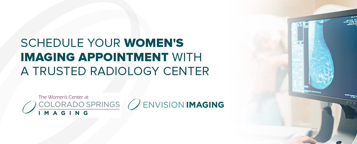
Schedule Your Women’s Imaging Appointment With a Trusted Radiology Center
If you’re looking for a caring and dependable place for women’s imaging, explore the services available from Envision Imaging or The Women’s Center at Colorado Springs Imaging. We prioritize delivering world-class women’s imaging by using the highest-quality scans and radiology services. We strive to enhance our patients’ health and lives by providing a pleasant imaging experience with compassionate care.
Those in the Colorado Springs area can book an appointment at The Women’s Center at Colorado Springs Imaging. Otherwise, locate the Envision Imaging center nearest you to schedule your women’s imaging test today.
Sources:
 Women’s imaging is image tests specifically for women’s health. While this may seem obvious, there is far more to this sector of radiology than that. Because women have different health concerns than men, they need image tests designed especially for them. From helping women catch diseases before they develop into larger issues to monitoring pregnancy, women’s imaging delivers this specialized care. This article will explore the field of women’s imaging, along with the main types of women’s imaging and where to find a dependable women’s imaging specialist.
 An ultrasound uses high-frequency sound waves to deliver real-time images of the inside of a patient’s body, including blood flow and the structure of internal organs. By using sound waves that can travel through soft tissue and fluids but bounce back upon hitting denser surfaces, an ultrasound can produce an accurate picture. Because the test does not use radiation, it is a safe technique to use during pregnancy.
Along with pregnancy imaging, ultrasounds can be used to diagnose a wide range of health conditions. For example, breast ultrasounds are used to detect breast tissue abnormalities or explore potential abnormalities found in a mammogram. With an ultrasound, a doctor may be able to determine whether a breast lump is a benign cyst or potentially a cancerous tumor. In addition, a breast ultrasound can help medical professionals target the correct area for a breast biopsy.
An ultrasound uses high-frequency sound waves to deliver real-time images of the inside of a patient’s body, including blood flow and the structure of internal organs. By using sound waves that can travel through soft tissue and fluids but bounce back upon hitting denser surfaces, an ultrasound can produce an accurate picture. Because the test does not use radiation, it is a safe technique to use during pregnancy.
Along with pregnancy imaging, ultrasounds can be used to diagnose a wide range of health conditions. For example, breast ultrasounds are used to detect breast tissue abnormalities or explore potential abnormalities found in a mammogram. With an ultrasound, a doctor may be able to determine whether a breast lump is a benign cyst or potentially a cancerous tumor. In addition, a breast ultrasound can help medical professionals target the correct area for a breast biopsy.
 Sources: