Considering that prostate cancer is the second most common type of cancer found in men, it is incredibly important for men to be aware of the disease. A prostate cancer diagnosis is not rare, and the illness can often be successfully treated when detected before it has the opportunity to spread. To catch prostate cancer early, men must take advantage of the screening and imaging tests available for diagnosing the disease.
To better inform patients about prostate cancer testing, we’ll discuss what prostate cancer is, the methods most medical professionals use to test for prostate cancer and where you can go for reliable prostate cancer imaging testing.
What Is Prostate Cancer?
Prostate cancer is a type of cancer that develops when the cells within the prostate gland begin to grow out of control. Because the prostate gland is part of men’s anatomy responsible for producing some of the fluid found in semen, prostate cancer is a disease found in males only. While younger men tend to have a smaller prostate gland, the gland tends to change and grow as a man ages.
The prostate is located beneath the bladder and in front of the rectum. Right behind the prostate gland are more glands called seminal vesicles that make the majority of the fluid for semen. The urethra, the tube that transports semen and urine out of the male’s body, runs through the center of the prostate gland.
Nearly all prostate cancers are adenocarcinomas, which means they develop from the gland cells that generate the prostate fluid added to the semen. However, other types of cancer can begin in the prostate, such as small cell carcinomas and neuroendocrine tumors. Other types of prostate cancer are extremely rare, so those who are diagnosed with prostate cancer almost certainly have an adenocarcinoma form.
While some prostate cancers can grow and spread rapidly, most progress slowly. In some cases, autopsy studies have found that older men who died of other causes had prostate cancer but didn’t know it because the disease didn’t impact their lives.
How Do Doctors Screen for Prostate Cancer?
Screening for a disease involves testing for it even if no symptoms or history of the illness are present. Because all men are at risk for prostate cancer, especially older men, getting screened is an important health care step for any man to take. For men not experiencing prostate cancer symptoms, the most common screening method is a prostate-specific antigen (PSA) blood test.
A PSA is a protein created by cells within the prostate gland. A blood test designed to assess PSA levels measures the concentration of PSA within a patient’s blood. Although there is no definitive cutoff number, the higher the PSA level in a man’s blood, the greater the chances he has prostate cancer.
Typically, men without prostate cancer have a PSA level under 4 nanograms of PSA per milliliter of blood (ng/mL), which means those with PSA test results above 4 ng/mL may require further testing. While the likelihood of a patient having prostate cancer decreases dramatically with a 4 ng/mL result, some doctors may request that those with lower PSA levels get additional testing. When done in tandem with other scans and tests, a PSA blood test can help medical professionals provide a more accurate prostate cancer diagnosis.
Imaging Tests Used to Diagnose Prostate Cancer
Along with a PSA blood test, imaging tests may be used to diagnose prostate cancer more decisively. Catching prostate cancer early through a variety of testing techniques can help a patient receive treatment before the problem grows. An imaging test of the prostate gland can give doctors a better grasp on the patient’s situation, direct the best ways to treat a patient and help health care professionals monitor the spread of the disease.
Below are the imaging tests most frequently used to detect prostate cancer:
1. Ultrasound
By using high-frequency sound waves to produce images of the inside of the body, a prostate ultrasound can give a doctor a clearer picture of the state of a patient’s prostate. A transrectal ultrasound is often the test administered for a patient with high PSA levels or other prostate cancer symptoms.
The images provided by a transrectal ultrasound are shown in real time and can reveal the development and structure of the prostate gland. This type of imaging may also be used during a prostate biopsy, which involves removing a sample of the prostate to test for cancerous cells. The ultrasound can guide the needles to the correct section of the prostate.
2. MRI
A magnetic resonance imaging (MRI) scan is one of the most common imaging tests because it can be an effective and painless way for doctors to assess the body’s soft tissues. An MRI scan uses powerful magnetic fields and radio waves to capture a detailed picture of the body’s tissue composition. These images can indicate whether there is an abnormal mass, such as a tumor, or another health issue present, which makes them helpful for detecting cancer.
Because an MRI generates internal images of specific areas, the test is extremely helpful for examining the prostate gland. An MRI scan is also a useful tool for medical professionals when they’re determining which treatment option would be best for a patient.
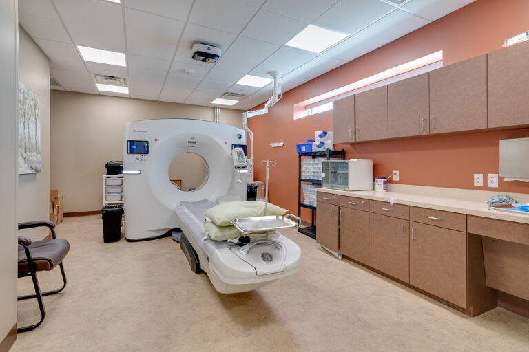
3. CT Scan
A computed tomography (CT) scan combines a series of x-ray views to provide an accurate look at the body’s bones and connected tissues. Medical professionals can view the images produced by CT technology either individually or as a 3D representation. A clear CT scan helps doctors assess the prostate for a diagnosis or monitor prostate cancer and treat it effectively.
Similar to an MRI, a CT scan produces images of the soft tissues inside the body. However, a CT scan also shows bones and can be used to track whether the prostate cancer has spread to any nearby areas or begun to grow on any of the surrounding organs.
4. Bone Density Scan
Prostate cancer has a tendency of spreading to the bones, so a bone density scan is a helpful tool for keeping tabs on the status of the disease. A bone density scan uses low-dose x-rays to generate a profile of the bone’s mineral makeup, which can help a doctor determine whether or not the cancer has spread. This vital information often influences a patient’s prostate cancer treatment plan.
By assessing the mineral density of the bones, a bone density scan can also indicate the strength of the patient’s bones. Patients who already know they have osteoporosis or a family history of the condition should especially consider receiving a bone density scan.
Schedule an Imaging Appointment at Envision Imaging or Colorado Springs Imaging
If you or a loved one needs an imaging test for diagnosing prostate cancer, book the appointment with a reliable radiology center like Envision Imaging or Colorado Springs Imaging. We put the needs of our patients first by providing the highest-quality imaging tests along with compassionate care. We offer exceedingly responsive and flexible scheduling options and immediate turnaround times for procedure results to meet patients’ needs.
To book your prostate cancer imaging test in Louisiana, Texas or Oklahoma, find the Envision Imaging office nearest you today. For patients in or around Colorado Springs, feel free to schedule your imaging appointment quickly and efficiently by contacting Colorado Springs Imaging.
Sources

 Considering that prostate cancer is the second most common type of cancer found in men, it is incredibly important for men to be aware of the disease. A prostate cancer diagnosis is not rare, and the illness can often be successfully treated when detected before it has the opportunity to spread. To catch prostate cancer early, men must take advantage of the screening and imaging tests available for diagnosing the disease.
To better inform patients about prostate cancer testing, we’ll discuss what prostate cancer is, the methods most medical professionals use to test for prostate cancer and where you can go for reliable prostate cancer imaging testing.
Considering that prostate cancer is the second most common type of cancer found in men, it is incredibly important for men to be aware of the disease. A prostate cancer diagnosis is not rare, and the illness can often be successfully treated when detected before it has the opportunity to spread. To catch prostate cancer early, men must take advantage of the screening and imaging tests available for diagnosing the disease.
To better inform patients about prostate cancer testing, we’ll discuss what prostate cancer is, the methods most medical professionals use to test for prostate cancer and where you can go for reliable prostate cancer imaging testing.

 Sources