 No one wants to hear they need to get a brain MRI. If your doctor ordered a head or brain MRI, we understand if you feel nervous for many different reasons. Even though an MRI machine looks intimidating, there is nothing to fear. Getting an MRI exam is a painless, safe and common procedure. For example, according to the International Society for Magnetic Resonance in Medicine, about 10 million patients get MRIs every year.
You may have some questions and concerns about MRIs. We hope to calm your worries and provide the answers you’re looking for. In this post, we’ll explore everything you need to know about a brain MRI to help you prepare and benefit from a life-saving diagnostic tool with as little stress as possible.
Jump to Sections:
No one wants to hear they need to get a brain MRI. If your doctor ordered a head or brain MRI, we understand if you feel nervous for many different reasons. Even though an MRI machine looks intimidating, there is nothing to fear. Getting an MRI exam is a painless, safe and common procedure. For example, according to the International Society for Magnetic Resonance in Medicine, about 10 million patients get MRIs every year.
You may have some questions and concerns about MRIs. We hope to calm your worries and provide the answers you’re looking for. In this post, we’ll explore everything you need to know about a brain MRI to help you prepare and benefit from a life-saving diagnostic tool with as little stress as possible.
Jump to Sections:
- What is a Head MRI?
- How Does a Head MRI Work?
- Why Would You Need a Head MRI?
- What Does an MRI of the Brain Show?
- How to Prepare for a Head MRI
- What to Expect With a Brain MRI
- How Long Does a Brain MRI Take?
- What Happens After a Head MRI?
- Schedule an Appointment With Us Today
What is a Head MRI?
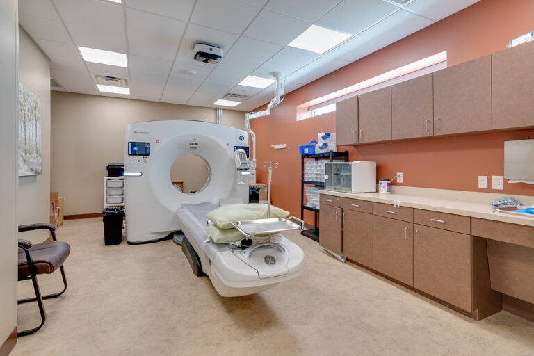
A head MRI is a noninvasive imaging test that creates detailed pictures of your brain and surrounding tissues. An MRI allows your doctor to see inside your brain to check for diseases or injuries without having to do surgery. Your doctor can use the images to make a diagnosis and recommend the best diagnosis treatment for your condition.How Does a Head MRI Work?
An MRI uses powerful magnets, radio waves and a computer to take images of your brain. MRI images are clearer and more precise than other forms of diagnostic imaging. Unlike CT scans and x-rays, an MRI does not use radiation.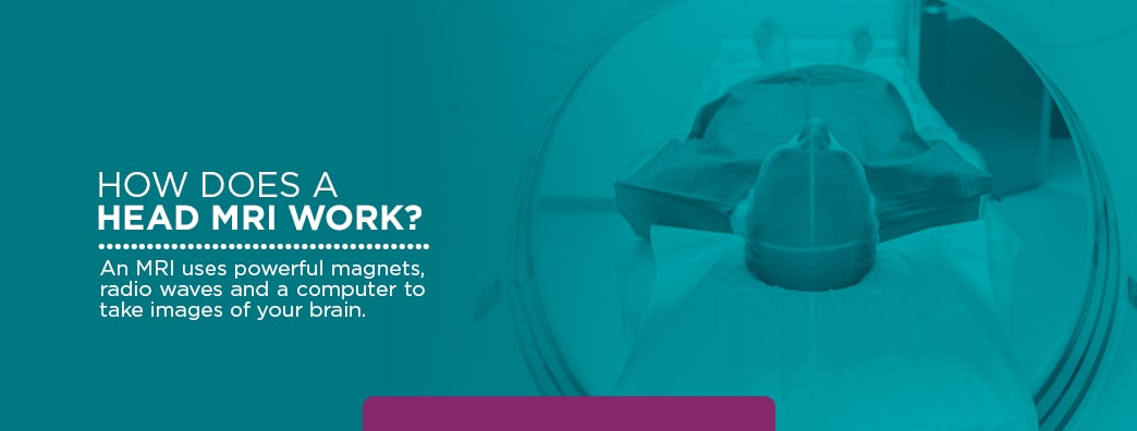 When your body is placed in an MRI scanner, it enters a strong magnetic field which causes your hydrogen protons to realign. This does not cause chemical changes to your body’s tissues. As the protons move back to their original alignment, they emit energy. The MRI machine captures this energy to create pictures. Radio waves transmit a signal to a computer which then processes MRI images.
Because an MRI provides clear pictures of soft tissues, it’s a reliable tool for diagnosing brain conditions. Soft tissues refer to muscles, fat, blood vessels, nerves and other tissues in the body. The human brain is almost 60 percent fat and around 75 percent water. Brain tissue is also made up of about 100 billion nerve cells. An MRI is the most sensitive imaging test for brain scans today.
When your body is placed in an MRI scanner, it enters a strong magnetic field which causes your hydrogen protons to realign. This does not cause chemical changes to your body’s tissues. As the protons move back to their original alignment, they emit energy. The MRI machine captures this energy to create pictures. Radio waves transmit a signal to a computer which then processes MRI images.
Because an MRI provides clear pictures of soft tissues, it’s a reliable tool for diagnosing brain conditions. Soft tissues refer to muscles, fat, blood vessels, nerves and other tissues in the body. The human brain is almost 60 percent fat and around 75 percent water. Brain tissue is also made up of about 100 billion nerve cells. An MRI is the most sensitive imaging test for brain scans today.
Schedule a Head MRI at Envision Imaging
Why Would You Need a Head MRI?
Your doctor might prescribe an MRI to look at “slices” of your brain so they can see what’s causing your symptoms. An MRI will help your doctor make an early diagnosis of a range of health conditions. You might get a head MRI exam so your doctor can check for any of the following:- Infections
- Stroke
- Brain tumors
- Multiple sclerosis
- Hemorrhage or bleeding in the brain
- Eye or inner ear disorders
- Pituitary gland disorders
- Blood clots
- Brain aneurysm or bulging of brain blood vessels
- Developmental abnormalities
- Spinal cord injuries
- Cysts
- Swelling
- Inflammation
- Hormonal disorders
- Hydrocephalus, or a buildup of fluid in the brain
- Vision problems
- Dizziness
- Seizures
- Chronic headaches
- Muscle weakness
- Muscles numbness or tingling
- Change in behavior or thinking
- Hearing loss
- Speaking difficulties
What Does an MRI of the Brain Show?
By looking at MRI images, your doctor can see details of blood flow and fluids surrounding the brain, which can help determine abnormalities in the brain relating to arteries and veins. An MRI brain scan also shows brain lesions. A brain lesion appears as a dark or light spot that does not look like normal brain tissues. Brain lesions may be present due to multiple sclerosis or as a result of an infection or a tumor. In general, a brain MRI will enable your doctor to examine blood flow and tissue health in the following brain structures.- Cerebrum: The cerebrum is the front part of the brain that involves movement, body temperature, touch, vision, hearing, reasoning, emotions and learning.
- Brainstem: The brainstem is the middle of the brain. This region involves eye and mouth movement, sensory messages, hunger, consciousness, cardiac function and involuntary muscle movements.
- Cerebellum: The cerebellum is the back of the brain which coordinates voluntary muscle movements and helps you maintain posture and balance.
How to Prepare for a Head MRI
You should not have to do too much to prepare for a head MRI. You may be able to eat, drink and take your medications as usual. However, if your doctor ordered a scan for other parts of your body, such as your abdominal region, they may instruct you not to drink or eat four to six hours before the test. Because an MRI machine is essentially a magnet, it can interact with metals, thus creating a blurry image. That’s why it helps to leave metal items at home before your appointment, or remove them before you enter the scanning room. You can prepare for your scan ahead of time by removing the following items from your body and pockets:- Body piercings
- Pens
- Jewelry
- Hearing aids
- Pins
- Hairpins
- Zippers or any metal clothing fasteners
- Removable dental work
- Kidney disease: If you have a history of kidney failure, kidney disease or liver disease, you may not be able to receive gadolinium, an intravenous contrast agent, which helps improve the accuracy of an MRI scan. Gadolinium increases your risk of nephrogenic systemic fibrosis, which is a disease that affects the skin and other organs.
- Pregnancy: There is a link between gadolinium and an increased risk of harm to the fetus. Therefore, doctors recommend women should not get an MRI with gadolinium anytime during pregnancy unless necessary.
- Claustrophobia: Claustrophobia is a fear of enclosed spaces. If you experience claustrophobia, you might speak with your doctor about taking anti-anxiety medication for the test. Many people worry about claustrophobia when it’s time for their MRI. Medical staff understand MRIs can seem frightening to individuals with anxiety or claustrophobia, but they are there to help you remain calm throughout the entire experience.
- Brain aneurysm clips, metal plates or any metal implants
- Artificial heart valve
- Pacemaker
- Inner ear implants
- Artificial joints
- Blood vessel stent
- Metal fragments in the body
- Any type of implantable pump
- Bullet wound
What to Expect With a Brain MRI
Knowing what to expect can help calm any of your anxiety about your MRI. Here are typical steps involved with getting an MRI.1. Remove Metal Materials
First, medical technologists will instruct you to remove and store any metal materials in a lockable storage area. You may wear comfortable clothes that do not contain metal or change into medical scrubs (top and pants) or a hospital gown once you get to the MRI center.2. Enter the Scanning Room
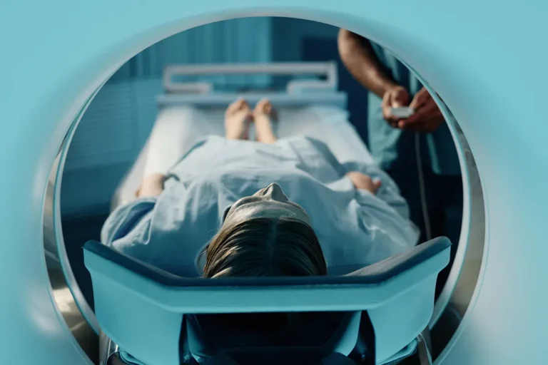
Medical staff will guide you into a special room that holds the MRI scanner. An MRI machine looks like a giant tube with openings at both ends. If you’ve experienced an MRI in the past, you may be expecting a tight tube. Newer machines are bigger and provide more space, which may help ease claustrophobia.3. Lie Down on the MRI Table
You’ll be asked to lie down on a table that slides into the tube. At Envision Imaging, you may choose an open MRI or MRI short-bore system, which may be best if you have claustrophobia. A short-bore system only scans the necessary part of the body, and allows the rest of the body to be outside of the magnet. For this type of scanner, only the upper part of your body would be inside the tube for a brain MRI. With an open MRI, all sides of the machine are open. The technologist will give you a pillow or foam block to help keep your head in the right position. If you feel cold, a technologist can cover you with a sheet to make you more comfortable.4. Get Contrast IV
Depending on your doctor’s orders, you may receive an injection of gadolinium before your procedure to make the pictures clearer. You might experience a flushing sensation or a metallic taste in your mouth for a few moments after the injection. Contrast agents rarely cause allergic reactions. Besides the minor pinch of having a small needle IV, you should not feel pain with the contrast agent. If you’re nervous about the contrast, speak with your doctor to ask if it’s needed or to discuss other options.5. Block Noises
MRIs produce loud noises. The technologist will provide you with headphones that play music during your scan. You’ll hear loud thumping, knocking and clicking sounds coming from the machine. These noises are normal and nothing to worry about.6. Stay Still
The most important thing you’ll need to do is to remain still throughout the exam, which ensures the images are clear and accurate. If you have trouble staying still or feel nervous about it, you may ask for a medication to help you relax. The technologist will be in another room to control the scanner, but will be able to see you the whole time through a window. The technologist will also provide instructions throughout the test, which you’ll hear through speakers in the scanner.7. Hold Your Breath
Your technologist may ask you to hold your breath during the exam for a few seconds, depending on the test. If at any time you feel anxious or want to stop the test, you can hit a call button on the machine and talk to staff through an intercom. The technologist will guide you every step of the way to make sure you are as comfortable as possible. You may ask your partner or a family member to be with you in the room to help you feel more relaxed.8. Remove Contrast IV
After the scan, the technologist will remove the contrast IV and help you off the table. Stand up from the table slowly to avoid lightheadedness or dizziness. If you took a sedative, you might need to rest until it wears off, and you should have someone to drive you home.How Long Does a Brain MRI Take?
An MRI typically takes between 30 and 60 minutes. The exam may take longer if the contrast is involved. Staff will let you know how long the exam should take once you arrive.What Happens After a Head MRI?
After the MRI, your technologist may ask you to wait to determine if they need to take more images. Unless your doctor says otherwise, you can go about your day and eat normally. There is no special care required after an MRI scan.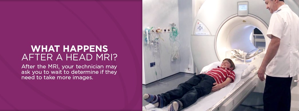 As you continue with your routine, a radiologist, which is a doctor with training to interpret MRI scans, will examine your MRI brain scan results and communicate with your doctor. You may wish to ask the staff how long it will take to hear back from your doctor about the results to help ease your mind.
If you notice any redness, swelling or pain from the IV site, let your doctor know, as this could be a sign of a reaction or infection. You may also feel some discomfort from having to lay still during the procedure, especially if you recently had surgery or an injury. Discuss this with the technologist ahead of time, and they will do all they can to reduce discomfort.
As you continue with your routine, a radiologist, which is a doctor with training to interpret MRI scans, will examine your MRI brain scan results and communicate with your doctor. You may wish to ask the staff how long it will take to hear back from your doctor about the results to help ease your mind.
If you notice any redness, swelling or pain from the IV site, let your doctor know, as this could be a sign of a reaction or infection. You may also feel some discomfort from having to lay still during the procedure, especially if you recently had surgery or an injury. Discuss this with the technologist ahead of time, and they will do all they can to reduce discomfort.
Schedule an Appointment With Us Today
Getting a brain MRI scan can seem intimidating, but we’re here to help you feel relaxed and in good hands. At Envision Imaging and Colorado Springs Imaging, our caring and compassionate staff genuinely want to make your experience as comfortable and convenient as possible while producing accurate images. We use the latest state-of-the-art imaging technology, ensuring fast turnaround rates, so your doctors can initiate treatment quickly. From the moment you schedule an appointment, we strive to accommodate your needs and make sure you have a positive, worry-free experience. To schedule an MRI, please contact one of our offices today. Sources
- https://medlineplus.gov/ency/article/003791.htm
- https://www.envrad.com/what-to-expect-when-getting-an-mri/
- https://www.ismrm.org/about/history-mission/
- https://www.ismrm.org/resources/information-for-patients/
- https://familydoctor.org/magnetic-resonance-imaging-mri/?adfree=true
- https://sma.org.au/resources-advice/injury-fact-sheets/soft-tissue-injuries/
- https://www.cancer.gov/publications/dictionaries/cancer-terms/def/soft-tissue
- https://www.ncbi.nlm.nih.gov/pubmed/20329590
- http://www.rehabchicago.org/the-human-brain/
- https://www.ncbi.nlm.nih.gov/books/NBK279302/
- https://www.ncbi.nlm.nih.gov/pmc/articles/PMC1121941/
- https://www.radiologyinfo.org/en/info.cfm?pg=headmr
- https://www.asnr.org/patientinfo/procedures/mri.shtml
- https://www.verywellhealth.com/what-to-expect-in-an-mri-3015134
- https://www.healthline.com/health/head-mri#uses
- https://www.hopkinsmedicine.org/health/treatment-tests-and-therapies/magnetic-resonance-imaging-mri-of-the-spine-and-brain
- https://www.ncbi.nlm.nih.gov/books/NBK279390/
- https://www.livestrong.com/article/262356-abnormal-brain-mri-results/
- https://www.mayoclinic.org/symptoms/brain-lesions/basics/definition/sym-20050692
- https://www.mayoclinic.org/diseases-conditions/nephrogenic-systemic-fibrosis/symptoms-causes/syc-20352299
- https://jamanetwork.com/journals/jama/fullarticle/2547756
- https://www.radiologyinfo.org/en/glossary/glossary1.cfm?gid=673
- https://www.medicalnewstoday.com/articles/323303.php
- https://www.radiologyinfo.org/en/info.cfm?pg=safety-mr
- https://www.envrad.com/your-experience/faqs/#mri
- https://www.self.com/story/what-to-expect-mri
- https://www.envrad.com/about-us/why-choose-us/
- https://www.envrad.com/locations/
- https://www.ncbi.nlm.nih.gov/books/NBK482487/